
Definition
Deformity of proximal femur with neck-shaft angle <110°
Issues
Limp / Trendelenberg gait
Stress fractures
Early osteoarthritis
Classification
Developmental
Acquired
- Rickets / Hypothyrodism / Renal osteodystrophy / Hyperparathyroidism
- Perthes disease / SCFE
- infection
- trauma / early closure physis
Dysplasia
- MED / SED www.boneschool.com/multiple-epithelial-dysplasia
- Achondroplasia www.boneschool.com/achondroplasia
- Fibrous Dysplasia www.boneschool.com/fibrous-dysplasia
Achondroplasia Multiple epithelia dysplasia
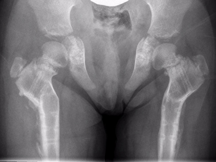
Developmental Coxa Vara
Epidemiology
Rare
Male = female
Bilateral in 1/3
Etiology
Unknown
Clinical
Abnormal gait
Trendelenberg gait
Reduced ROM
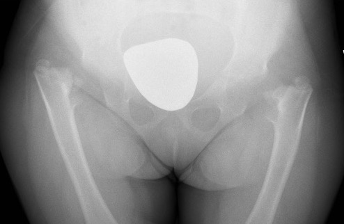
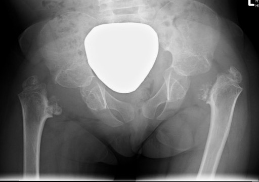
Xray
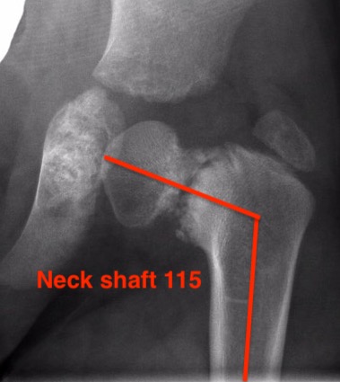
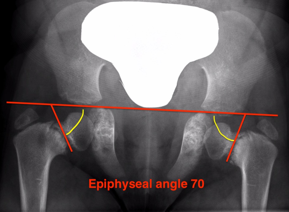
| Varus femoral neck | Inverted Y | Hilgenreiner's epiphyseal angle |
|---|---|---|
|
Neck-shaft angle < 125°
Normal is 150° in infant |
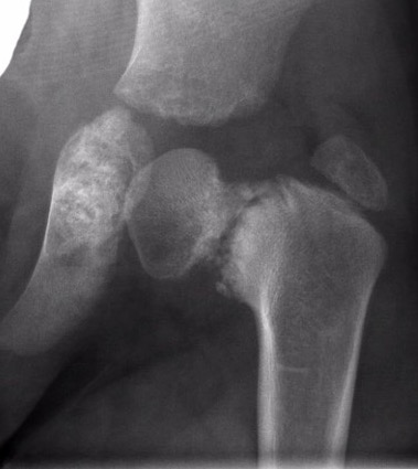
Inferior sclerotic metaphyseal triangle
Pathognomonic of developmental |
Angle between Hilgenreiner's & physeal line
Normal < 25°
|
Management
Management based on Epiphyseal Angle
| < 45 degrees | 45 - 60 degrees | > 60 degrees |
|---|---|---|
|
Unlikely to progress
|
Observe | Surgery |
|
Observe
|
Surgery if progresses |
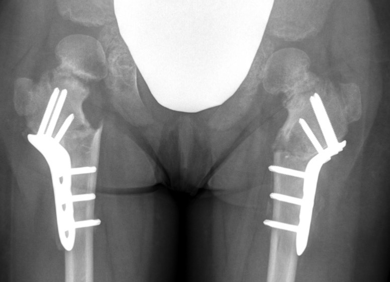
Operative management
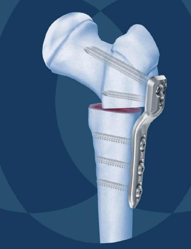
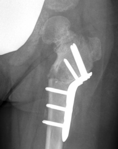
Valgus derotation subtrochanteric osteotomy
- overcorrect to 150˚
- epiphyseal angle < 40o
- correct anteversion to 10o
Results
- systematic review of proximal femoral osteotomy in 192 hips with coxa vara
- success rate was 89%
- loss of correction 11%)
- deep infection 1%
- revision surgery 9%
Technique
Synthes Pediatric Proximal Femur Offset Plate Technique PDF
POSA proximal femoral osteotomy for coxa vara video
Lateral approach
- elevate / L shaped detachment of vastus lateralis
- mark distal and proximal with drill hole for rotation
- K wire in central head
- sub-trochanteric osteotomy with saw
- application of 150o Synthes offset locking plate
- internal rotation of about 20° at time of osteotomy
May require
- adductor tenotomy
- femoral shortening
- greater torchanter transfer
Complications
Loss of correction - related to undercorrection
Premature physeal closure - related to increased pressure
Greater trochanter overgrowth - associated with premature physeal closure
Acetabular dysplasia - associated with premature physeal closure and undercorrection