
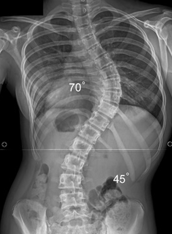
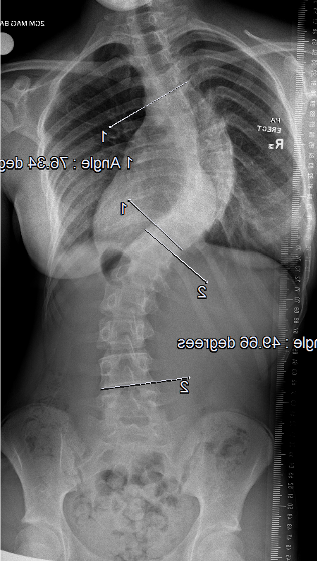
Definition
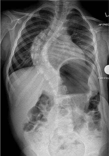
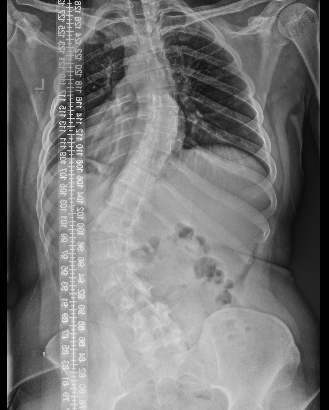
Lateral curvature of the spine
- > 10o coronal plane deformity with vertebral rotation
- over the age of 10
Epidemiology
Prevalence dependent on the size of the curve
- > 10o - 2 - 3%
- > 30o - 0.3%
- > 40o - 0.1%
Etiology
Unknown
Genetic / increased incidence in affected relatives
- mother with scoliosis 10% chance for female child
- sister with scoliosis 20% chance for female child
- mother and father with scoliosis 80% chance for female child
Xray
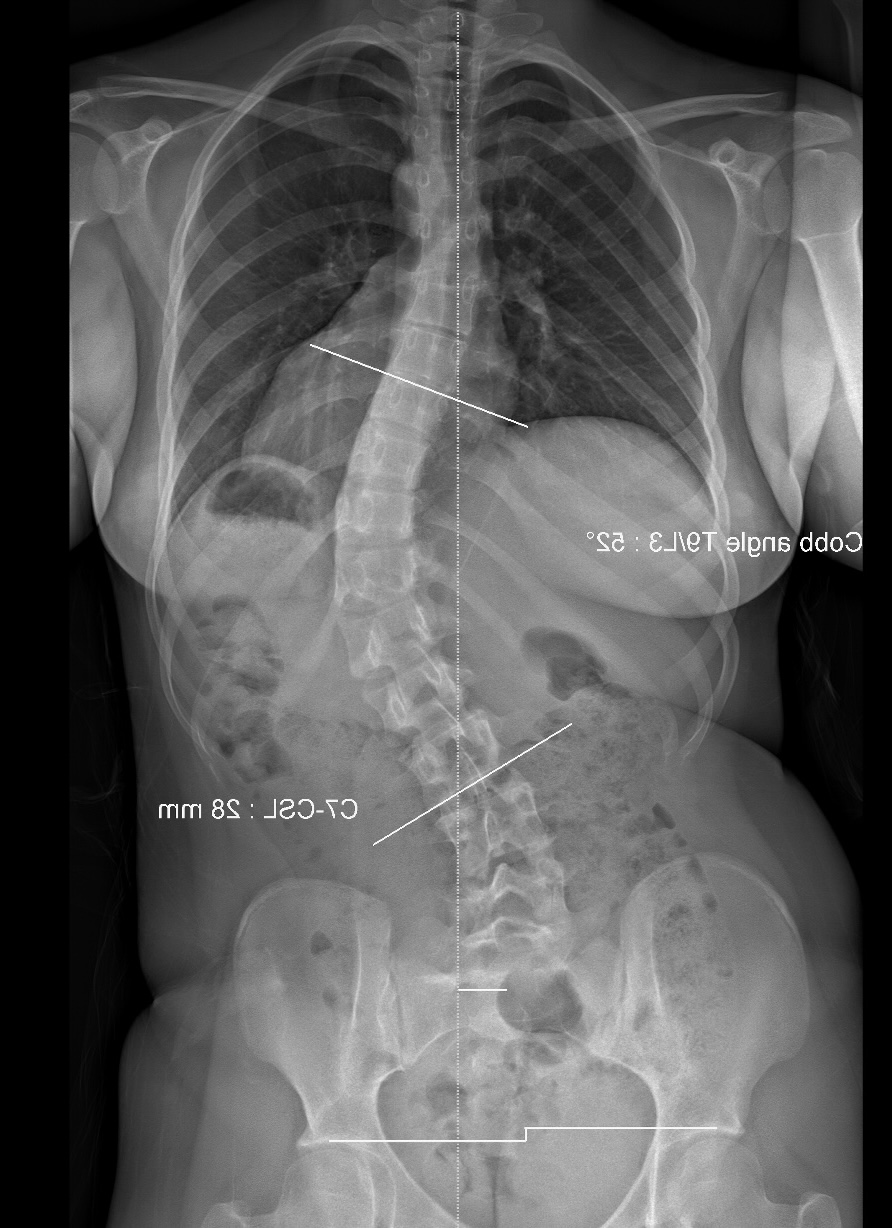
Standing AP or PA films of whole spine including the iliac crests
- levoscoliosis: curvature towards the left
- dextroscoliosis: curvature towards the right
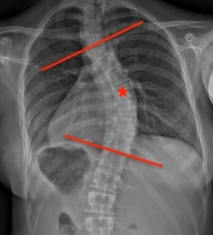
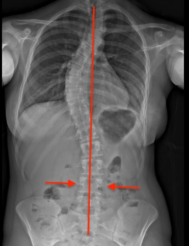
| End vertebrae | Apical vertebra | Stable vertebra |
|---|---|---|
|
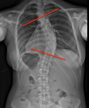
Last vertebra that tilts into the concavity of the curve
Form basis of Cobb Angle |
Center of the curve Furtherest horizontal deviation from center Most rotated
|
Lowest vertebrae bisected by the central sacral line
Lowest vertebrae instrumented in surgery |
 |
 |
 |
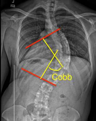
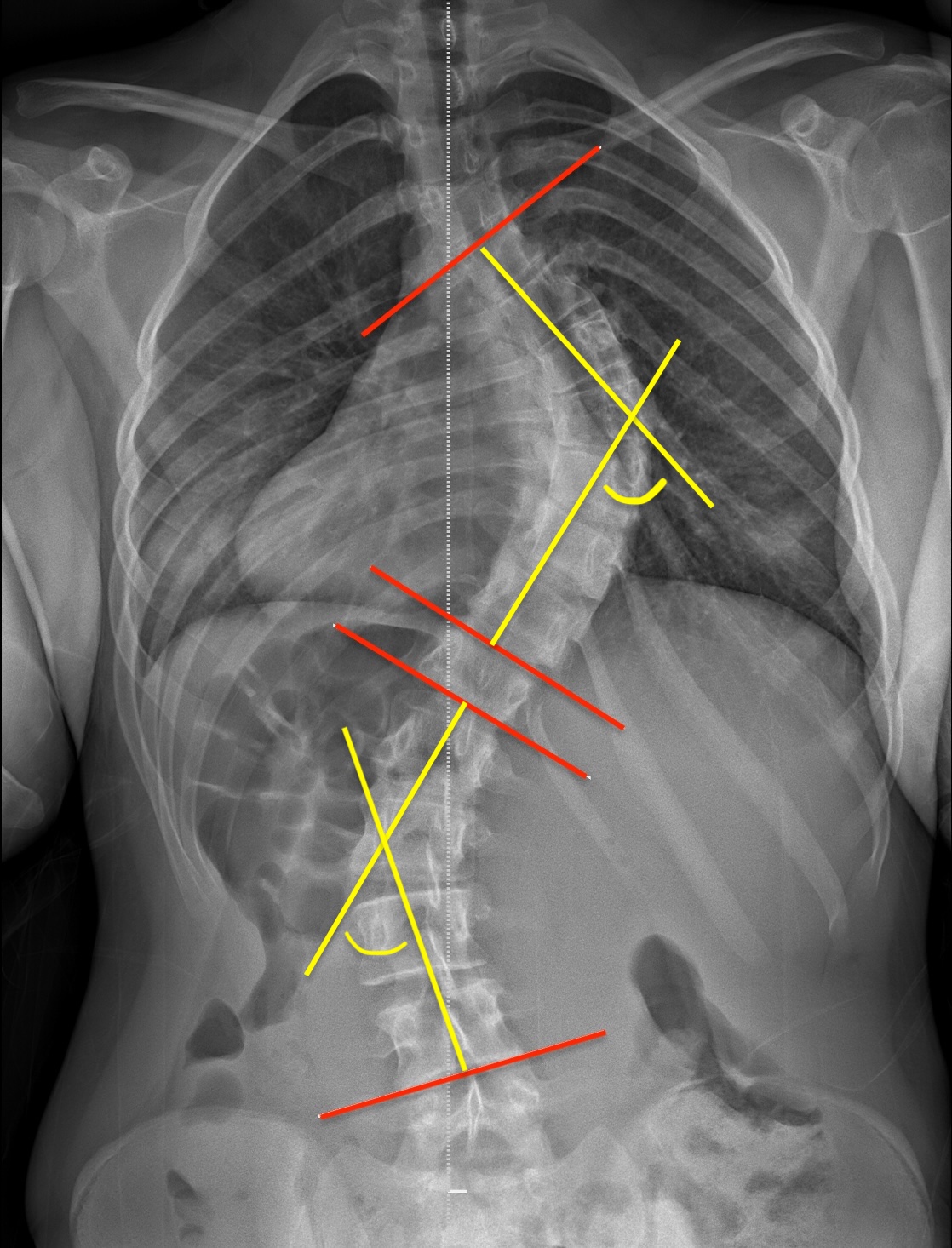
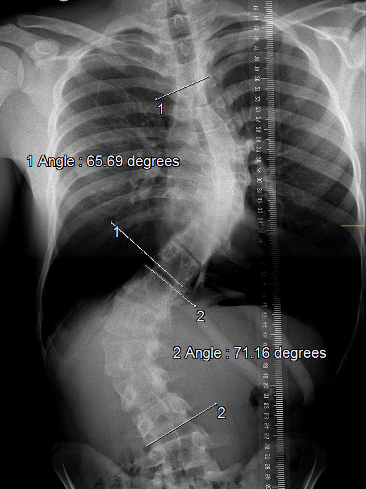
Cobb Angle
Lines drawn upper plate of upper end vertebra and lower plate of the lower end vertebra
- perpendiculars to these lines
- Cobb angle is the intersection angle
Lenke classification
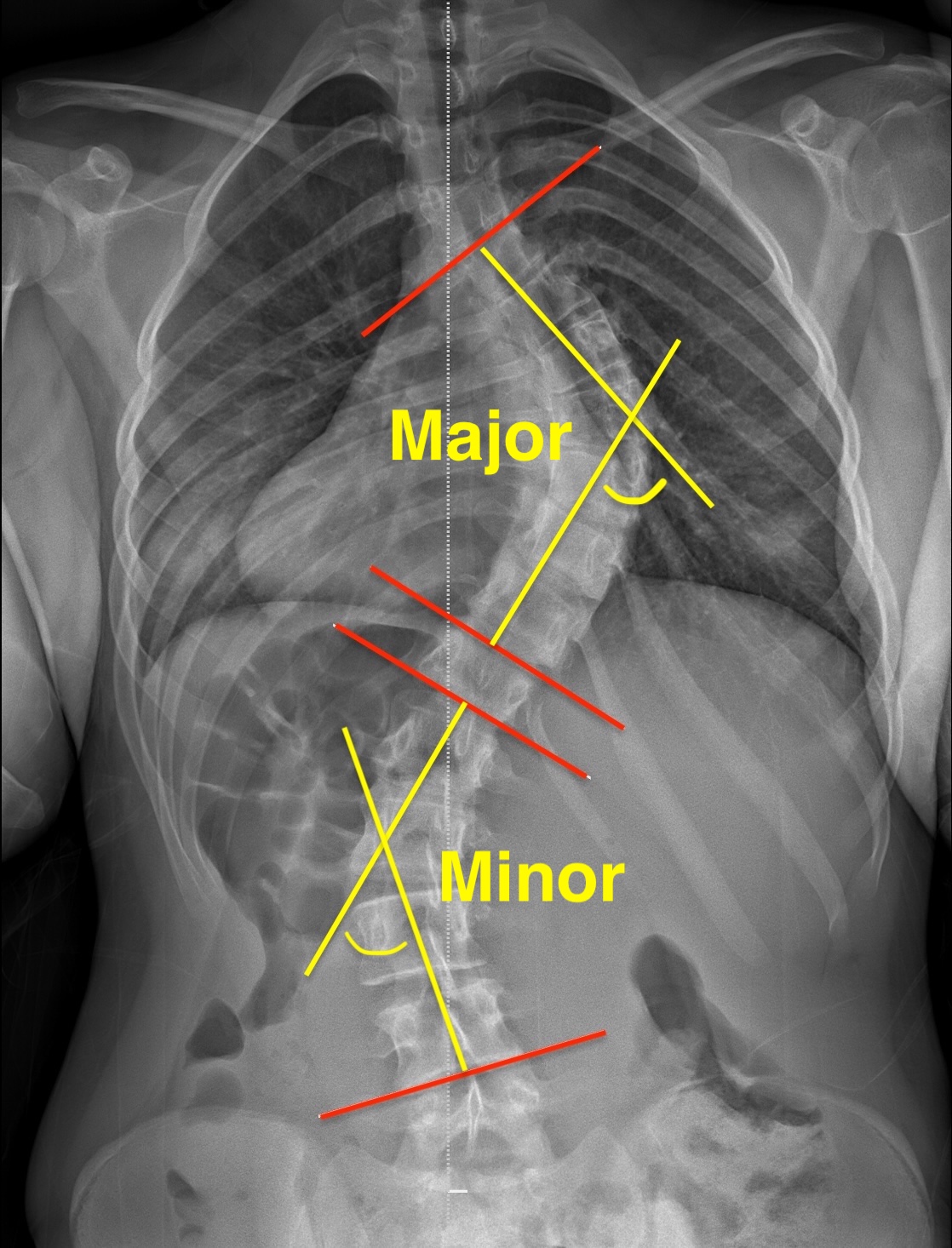
Six curve types based upon location of major curve / largest Cobb angle
Divide spine into 3 segments
- proximal thoracic - apex vertebrae T3/4/5
- main thoracic - apex vertebrae between T6 and T12
- thoracolumbar / lumbar - thoracolumbar apex between T12/L1, lumbar apex between L1 and L4
Major curve - the largest Cobb angle, only main thoracic or thoracolumbar / lumbar can be major curve
Minor curve - structural: > 25 degrees on lateral bending xrays
| Type 1: Main Thoracic | Type 2: Double Thoracic | Type 3: Double Major |
|---|---|---|
|
Structural curve main thoracic region
|
Major structural curve main thoracic region Minor structural proximal thoracic curve |
Major structural curve main thoracic region Minor structural thoracolumbar / lumbar curve
|
|
 |
 |
| Type 4: Triple major | Type 5: Thoracolumbar / Lumbar | Type 6: Thoracolumbar/lumbar - Main thoracic |
|---|---|---|
|
Major structural curve main thoracic region or Main structural thoracolumbar / lumbar curve Minor structural proximal thoracic curve |
Major structural thoracolumbar / lumbar curve |
Major structural thoracolumbar / lumbar curve Minor structural main thoracic curve |
|
 |
|
| Lumbar spine modifier (A,B,C) | Sagittal thoracic modifier (-, N, +) |
|---|---|
|
Center sacrum vertical line (CSVL) A: Between pedicles apical lumbar vertebrae B: Touches pedicle apical lumbar vertebrae C: Doesn't touch pedicle |
Side profile of the thoracic spine - hypokyphotic (-) - normal (N) - hyperkyphotic (+)
|
Lateral xray
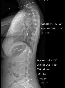
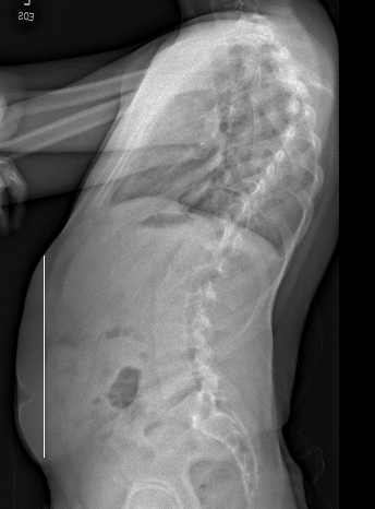
Measure the kyphosis and lordosis via Cobb method
- want to correct this intra-operatively
- usually need to recreate thoracic kyphosis
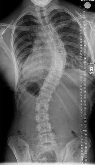
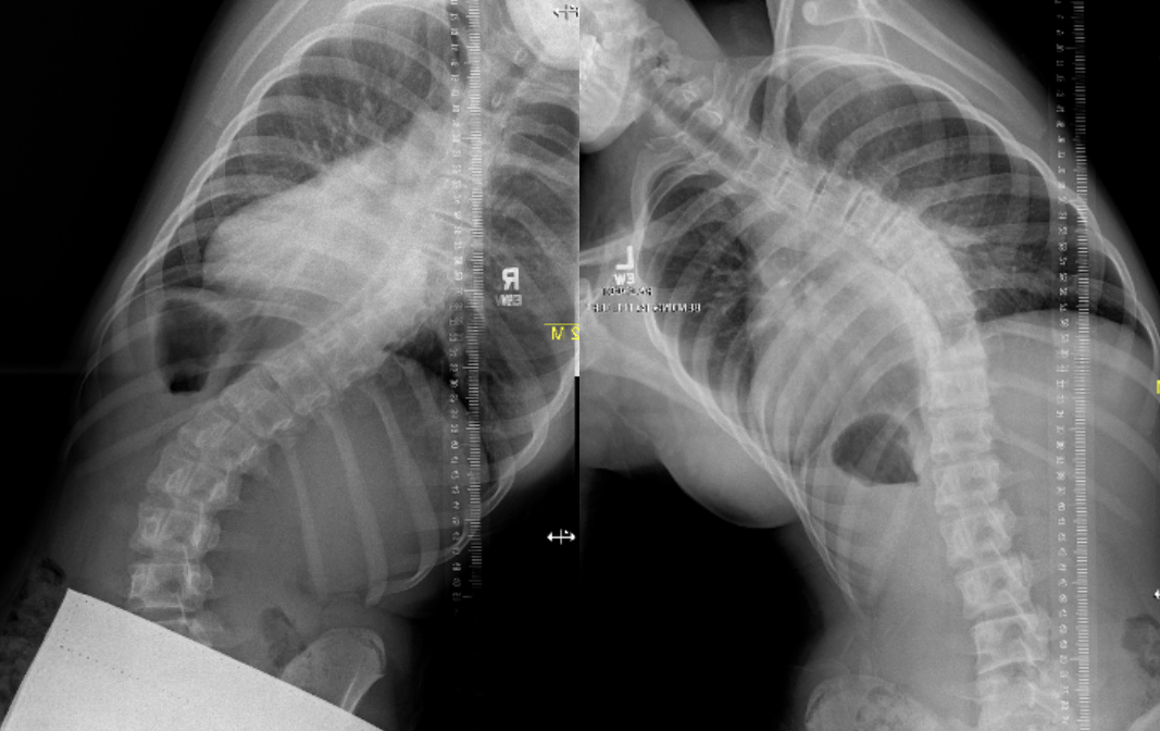
Lateral Bend Films
Push prone
- supine with maximal voluntary bend
- differentiates structural from compensatory curves
Progression of curve
Definition
Absolute increase in Cobb angle of 10o or 5o over two consecutive visits
Factors related to progression (MR Sex MAP)
Magnitude: curve > 20o
Risser: 0 or 1
Sex: Female
Menarche: premenarche
Age: < 12
Pattern: Thoracic & double curves highest risk
- systematic review of factors affecting curve progression in 8000 patients
- age < 13, peak height velocity, Risser < 1, Cobb angle > 25o , thoracic single or double curves
Peak height velocity (PHV)
Most important factor
- adolescent growth spurt generally over 2 years
- girls 8 cm / year, boys 9.5 cm / year
- before menarche
- Risser 0
- open triradiate cartilage
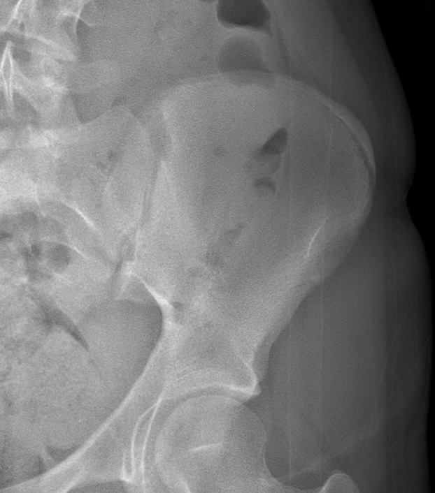
Risser sign
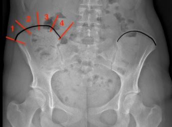
Ossification of iliac apophysis from lateral to medial
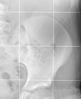
- Grade 0: no apophysis (mean age 10)
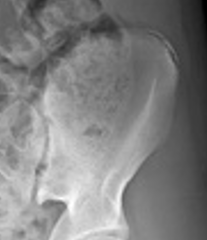
- Grade 1: lateral apophysis appears after menarche, < 25%
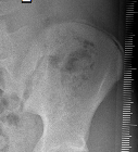
- Grade 2: 25 - 50%
- Grade 3: 50 - 75%
- Grade 4: > 75 - 100% but not fused to ilium
- Grade 5: fusion of apophyseal cap to ilium / no open growth plates long bones (mean age 16)
- review of 600 xrays
- Risser 0: mean age 10
- Risser 5: mean age 16
- females reach each stage sooner
- typically 1 year between stages
| Risser 0 | Risser 1 - 2 |
|---|---|
 |
 |
| Risser 2/3 | Risser 4 |
|---|---|
 |
 |
Natural History of Scoliosis
Back Pain
Impaired pulmonary Function - curves of > 80o
- 50 year follow up of 117 untreated scoliosis patients
- compared to age matched cohort
- increased risk of shortness of breath with curves of > 80o
- increased risk of back pain, but majority minor
Screening
US preventative services taskforce JAMA 2018
- no evidence for benefit of screening for idiopathic scoliosis
- evidence that bracing may slow curve progression
- no evidence this improves long term health outcomes
Examination
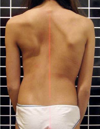
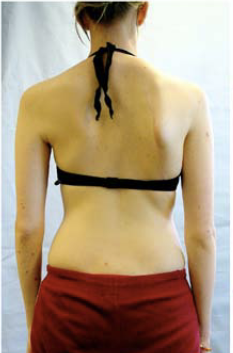
From: Weiss et al Scoliosis 2007
Typical curve
- the right shoulder is raised
- the right scapula is prominent
- the loin creases are asymmetrical / lumbar fullness
- the pelvis is level
- there is flattening of the normal thoracic kyphosis
- there is a normal lumbar lordosis
- on forward bending, there is a (mild/moderate/severe) (well rounded/angular) rib hump
Adam's forward bend test / rib hump
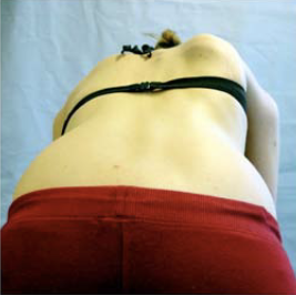
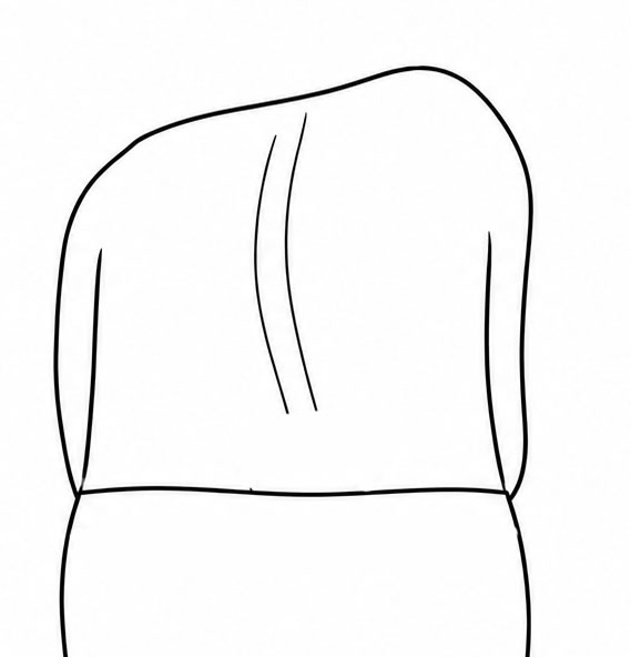
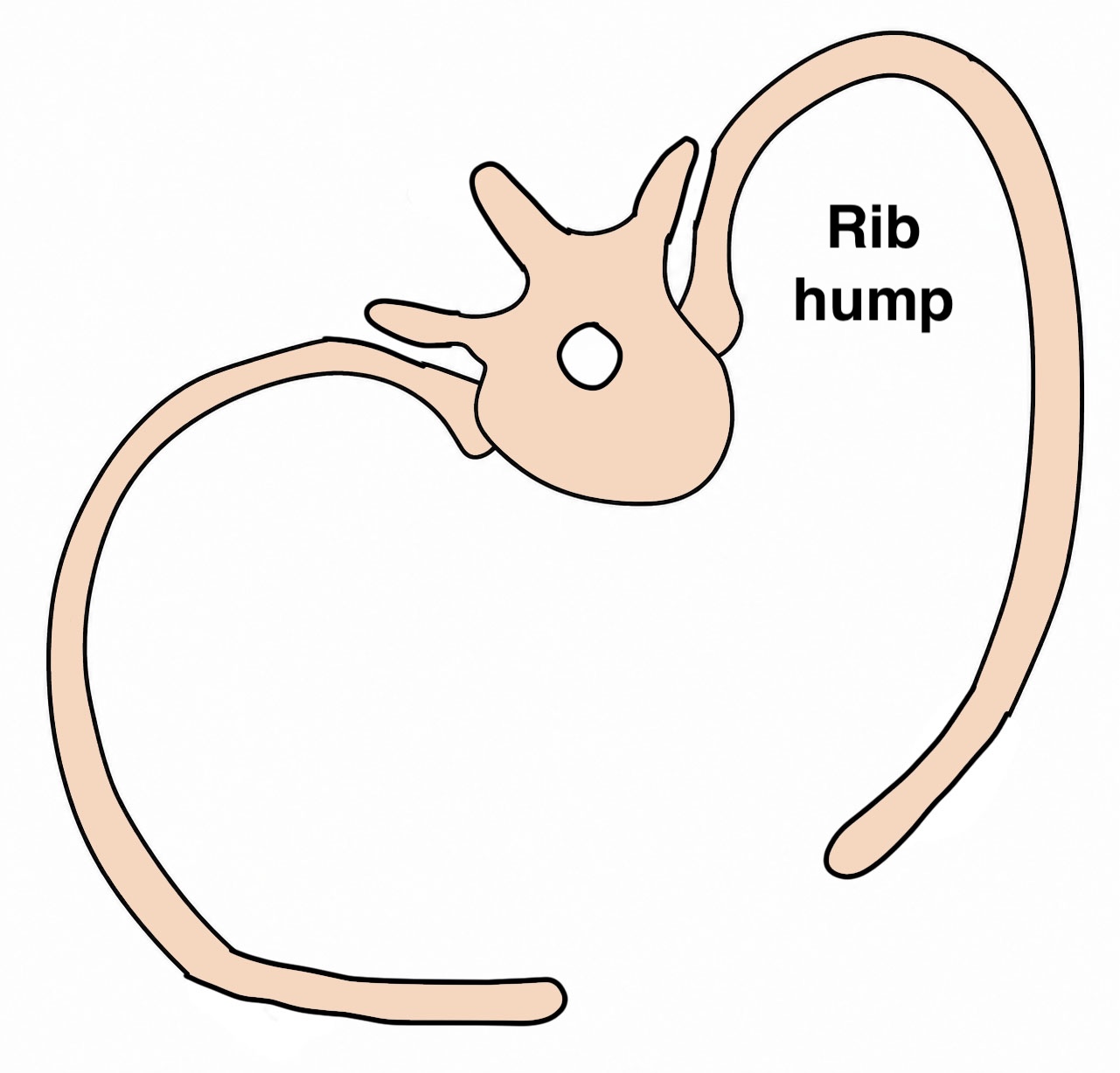
From: Weiss et al Scoliosis 2007
Hands together & bend forwards to touch floor
- mild / moderate / severe rib hump
- measure rib hump with scoliometer / inclinometer
- < 5o tilt = < 30o rotation
- > 7o tilt = > 30o rotation
Neurological assessment
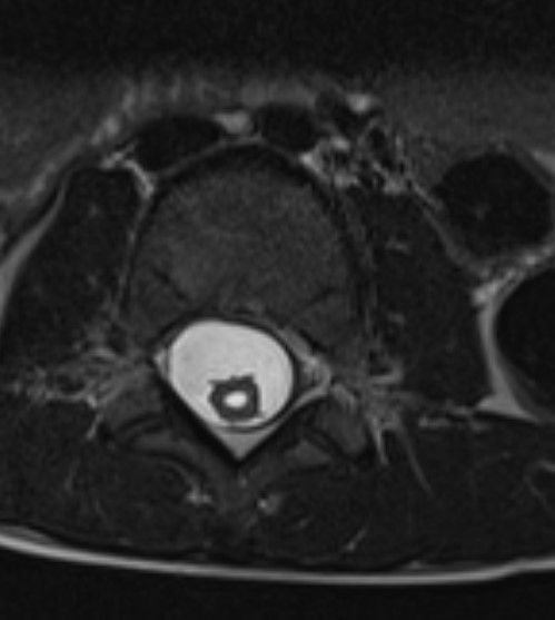
- scoliosis + °Abdominal reflexes & °Axillary sensation - syrinx till proven otherwise
- abdominal reflexes disappear during teens
Exclude non idiopathic
- Marfan's / Neurofibromatosis / Skeletal Dysplasia / spinal dysraphism
- leg length discrepancy
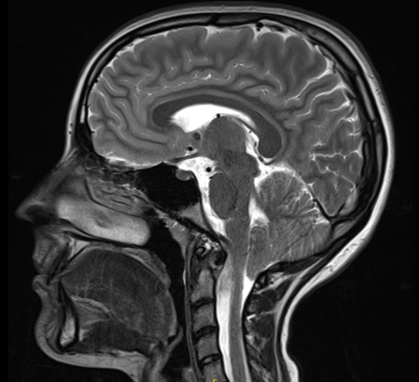
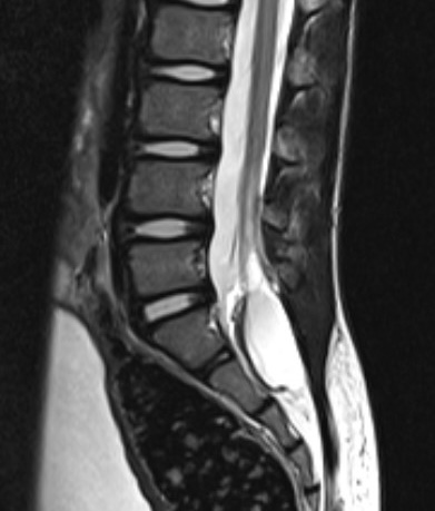
MRI
Issues
| Chiari malformation | Syrinx | Cord tethering |
|---|---|---|
 |
 |
 |
Incidence
Singhal et al Eur Spine J 2013
- preoperative MRI in all 206 adolescent idiopathic scoliosis undergoing surgery
- abnormalities in 10% of all patients
- 5% of all patients underwent a neursurgical procedure prior to scoliosis surgery
Indications
Left thoracic curves
Rapidly progressive curves
Neurological symptoms
Severe back pain