
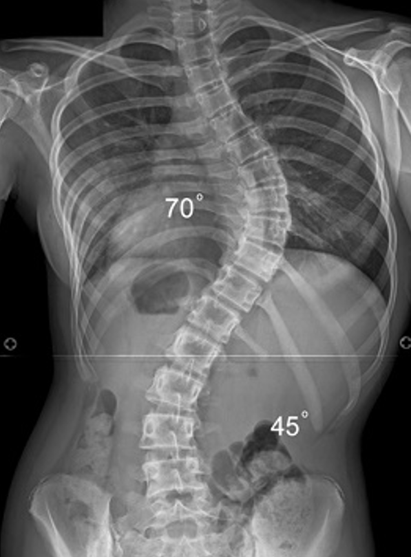
Curve Progression
Typically thought that curves > 45 - 50o continue to progress after skeletal maturity
Lonstein and Carlson JBJS Am 1985
- 727 patients with scoliosis followed to skeletal maturity
- 23% progressed
- correlation to magnitude curve / chronological age / Risser sign / menarche
Weinstein and Ponseti et al JBJS Am 1983
- 102 patients followed for 40 years
- 68% of curves progressed after skeletal maturity
- curves < 30o did not progress
- thoracic curves > 50 - 75o progressed most ( 1o/ year )
Options
| Cobb Angle | High growth potential / Risser 0 - 2 | Low growth potential |
|---|---|---|
| <20° | Observe | Observe / discharge |
| 20°-30° | Observe / brace | Observe |
| 30°-45° | Brace | Observe |
| >45° | Surgery | Surgery / observe |
Exercises
Dimitrijevic et al Eur Spine J 2022
- systematic review of Schroth method and core stabilization exercises
- positive effect on Cobb angle / trunk rotation / QOL
Bracing
Indications
Typically prevent curve progression / will not correct or reverse curve
1. Risser 0-2 (growth potential)
2. Curve >30o adolescent
3. Curve >25o with progression (5o in six months)
4. Willingness to comply - must wear 18 - 23 hours a day until skeletal maturity
Types
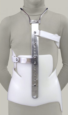
| Milwaukee Brace / CTLSO | Boston brace / TLSO |
|---|---|
|
For curves with apex above T8 - pelvic piece over iliac crests connected to neck piece - thoracic and lumbar pads |
For curves with apex under T8 |
 |
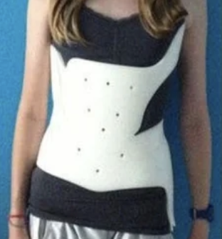 |
Results
Bracing
- systematic review of full time braces in adolescent idiopathic scoliosis
- good evidence that braces prevent curve progression
Night time bracing
Kodra et al J Pediatr Soc North Am 2026
- systematic review of Providence night time bracing
- good evidence of prevention of curve progression
- likely similar efficacy to full time bracrs
Operative Management
Indications
Typically thought that curves > 45 - 50o continue to progress after skeletal maturity and require surgical intervention
Indications for surgery
- Cobb > 45 - 50o
- progression of curve
- significant cosmetic deformity
- significant coronal or sagittal imbalance
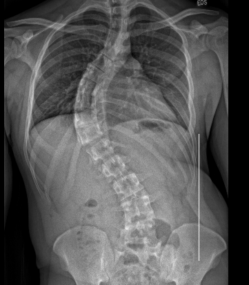
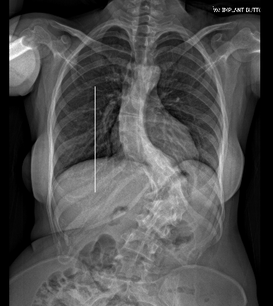
Principles
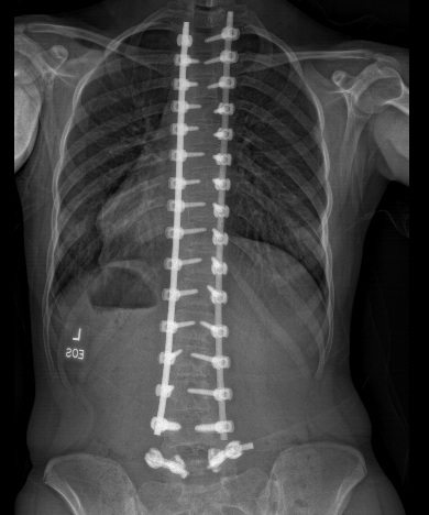
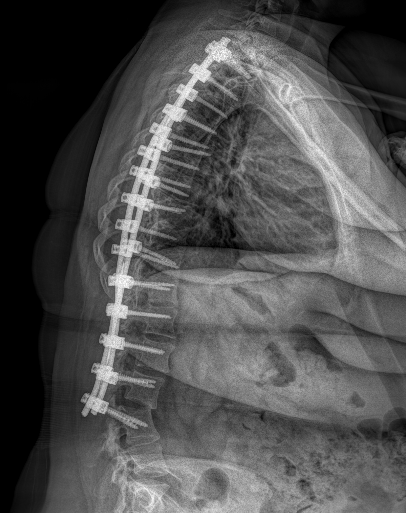
Obtain solid arthrodesis with correction of deformity and coronal / sagittal plane balance
Fuse minimal segments while obtaining adequate correction
- involving lumbar segments associated with early degeneration of unfused segments
- risk back pain
Options
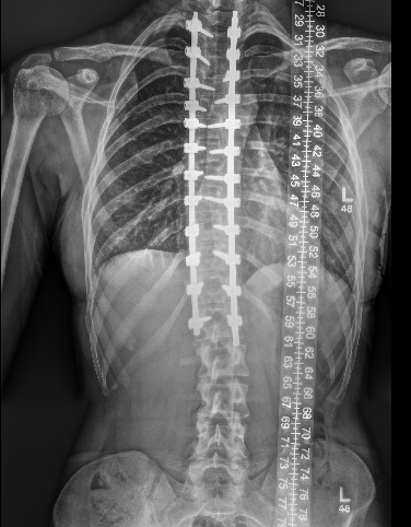
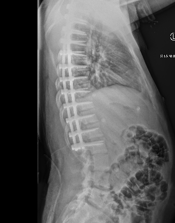
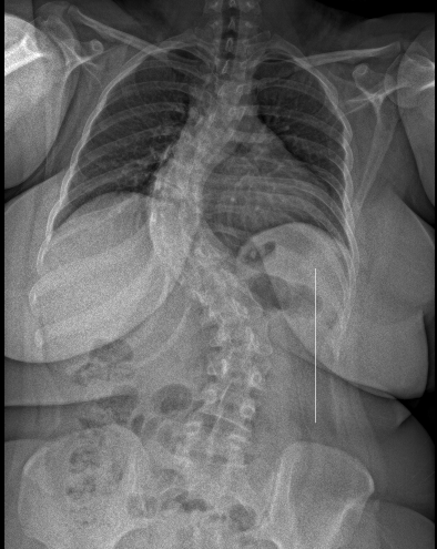
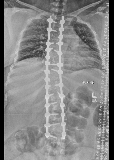
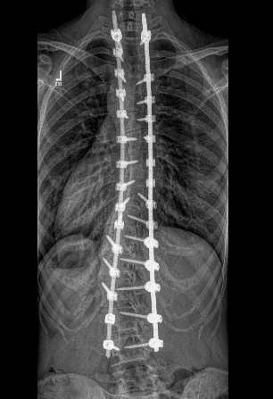
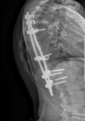
Posterior instrumented fusion
Anterior instrumented fusion
Anterior vertebral body tethering
Posterior Instrumented fusion
Instrumentation
Multisegmental hook and pedicle screw systems
- compression / rotation / distraction
- correction in the coronal and sagittal planes
Selective versus non selective thoracic fusion in Lenke Type I Main Thoracic curve
Selective thoracic fusion - avoid lumbar curve fixation
- systematic review of selective v non selective thoracic fusion Lenke Type 1 C
- shorter operative times with selective
- improved corrections with non selective
- no difference in QOL measures
- 106 patients Lenke Type 1C to 4C
- treated with selective thoracic fusion
- adequate correction if lumbar curve < 45° / bending lumbar Cobb angle < 25°
Low versus high density pedicle screw
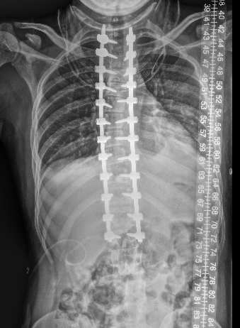

- systematic review of 24 cases and 2000 patients
- some better radiographic outcomes with high density screw use
- no difference in QOL
- increased cost and operative time with high density screw use
Hybrid (screw / hook) constructs versus all screw
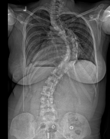
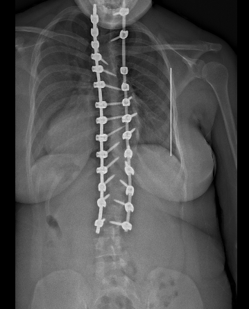
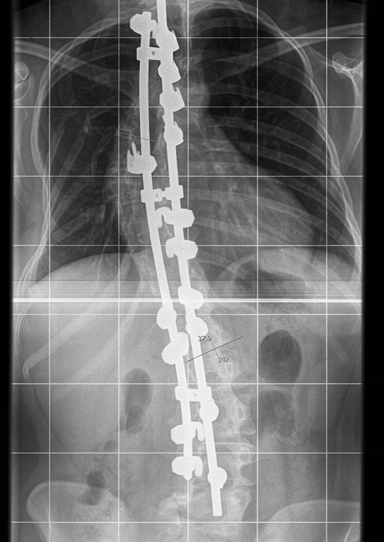
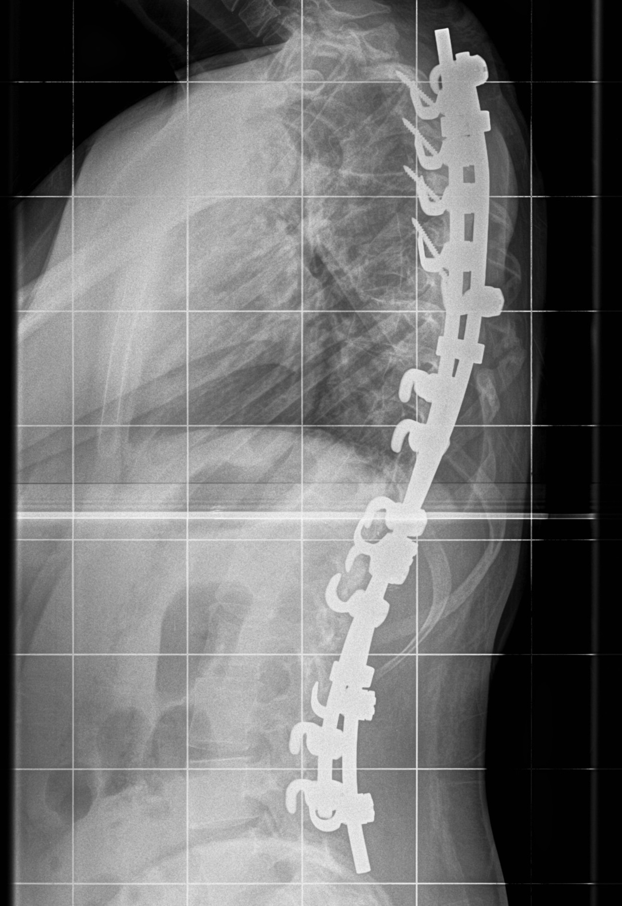
- systematic review of hybrid versus all screw constructs
- 28 studies and 3400 patients
- lower complication and reoperation rate with all screw constructs
- better coronal plane correction with all screw
- better sagittal plane correction with hybrid
Navigation
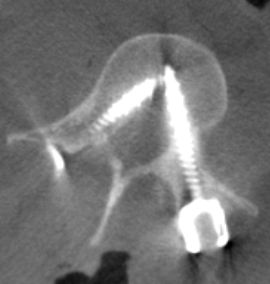
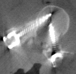
- systematic review of pedicle screw fixation for AIS
- free hand versus navigation techniques
- lower breach rate with navigation techniques
- longer operative times with navigation techniques
- no difference in complication rates
Open posterior fusion versus MIS (minimally invasive surgery)
Yang et al J Orthop Surg Res 2022
- systematic review of open v MIS pedicle screws for idiopathic scoliosis
- longer operative time with MIS
- reduced blood loss / reduced pain / reduced postoperative pain with MIS
Technique
AO surgery foundation posterior instrumented fusion Lenke Type 1
AO surgery foundation pedicle screw insertion technique by level
POSNA posterior instrumented fusion video
Spinal cord monitoring
Highly accurate at detecting spinal cord injury
- affected by other factors
- anesthesia: need total intravenous anesthesia as muscle relaxants / inhalation anesthesia affect readings
- also affected by: hypothermia, blood loss, low blood pressure
| Somatosensory evoked potentials (SEPs) | Transcranial motor evoked potential (TcMEP) |
|---|---|
|
Electrodes placed upon limbs - median nerve / wrist, tibial nerve / popliteal fossa - stimulate a waveform and measure in the cortex - measure baseline - concern with reduction in wave amplitude > 50% |
Electrodes placed on scalp - electrical stimulation measured in distal muscles |
Technique
- 4 Poster Bed - protect eyes, knees, elbows
- no pressure on abdomen / reduce venous bleeding
- midline incision / posterior approach
- divide thoracolumbar fascia midline
- split apophysis with knife (if present)
- subperiosteal elevation strap muscles
- expose facet joints and transverse processes
- don't go between transverse processes laterally as nerve roots here
- insert pedicle screws
- rods prebent in sagittal plane
- correct coronal malalignment and rotation as able
- decorticate lamina, add bone graft along each side
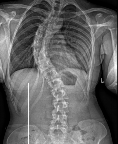
Levels
Lenke Type 1: Selective versus non selective thoracic fusion
Lenke Type 2 / Double Thoracic:) Posterior stabilization to T2 to stabilize / level shoulders
Lenke Type 3 (Double Major) / Type 4 (Triple Major) / Type 6: Long posterior instrumented fusion
Lenke Type 5 (Thoracolumbar / Lumbar): Lumbar spine fusion
Anterior fusion
Results
Benites et al Neurosurg Rev 2025
- systematic review of anterior v posterior instrumentation
- longer operative times for anterior instrumentation
- similar blood loss and correction
- fewer instrumentated vertebrae with anterior
Anterior vertebral body tethering
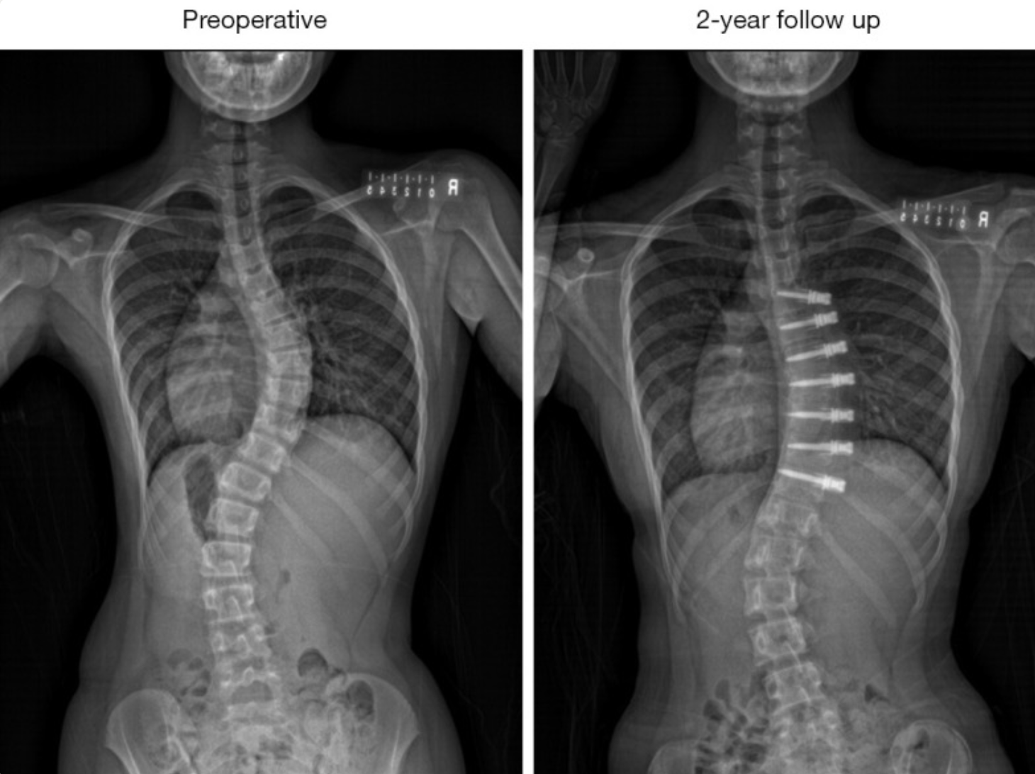
From: Wong et al J Spine Surg 2024
Concept
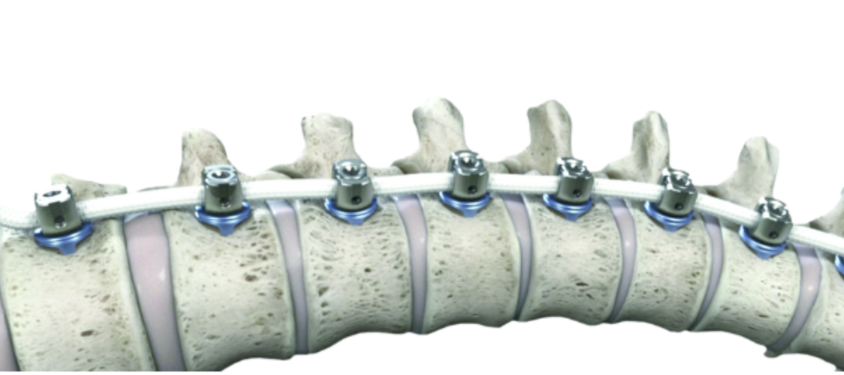
Youtube Zimmer Biomet Tether animation video
Non fusion method / growth modulation
- limit the growth of spine on one side
- allows the curve to progressively correct
Thoracoscopy used to place implants into vertebral body
Strong cords link implants and prevent growth
Results
- systematic review of 9 studies and 200 patients mean age 12
- overcorrection 14%
- mechanical complications 28%
- pulmonary complications atelectasis / pleural effusion 10%
- tether revision 8%
- revision to spinal fusion 8%
Varona et al Spine Deform 2025
- vertebral tethering in 156 patients
- best outcomes and lowest revision rate in Lenke Type 5 curves
Complications
Overall
Kwan et al BMC Musculoskeletal Disord 2021
- 1057 cases of surgery for AIS single center
- 87% female, mean age 16, with 47% Lenke 1 curve types
- mean operative time 2 hours
- mean blood loss 1 liter
- 5% transfusion rate
- superficial infection 1%
- deep infection 0.2%
- neurological injury 0.5%
- 2 superior mesenteric artery syndrome
- one massive blood loss
Revision
Dong et al Global Spine J 2024
- 1800 patients
- revision rate 2.8%
- malposition implants / infection / spinal decompensation / proximal junctional kyphosis
- revision rate lower with posterior versus anterior approach
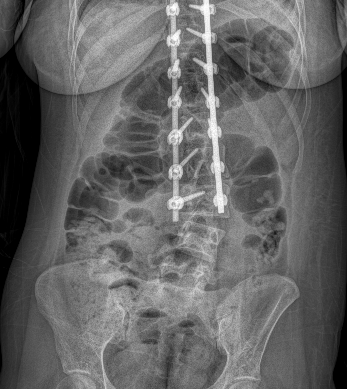
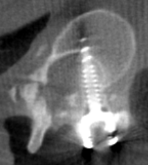
Superior mesenteric artery (SMA) syndrome
Scoliosis correction narrows distance between aorta and SMA
- compresses duodenum
- vomiting / abdominal pain / tenderness / distention
- more likely with low BMI and sagittal kyphosis
- diagnosis with oral contrast imaging
- attempt nonoperative with nasogastric tube
- may need duodenojejunostomy (form a junction between the jejunum and the duodenum)
Spinal decompensation / adding on
Coronal plane malalignment after scoliosis surgery
Proximal junctional kyphosis
Ji et al J Orthop Surg Res 2024
- systematic review
- overall incidence 19%
- one quarter with Lenke Type 5 curves
Zhong et al World Neurosurg 2019
- systematic review
- most common with larger thoracic kyphosis / larger lumbar kyphosis
Back pain
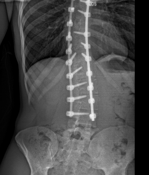
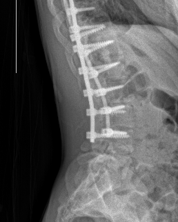
Lumbar adjacent disc degeneration / adjacent segment degeneration
- degenerative disc disease (DDD) below lowest instrumented vertebrae
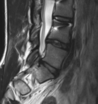
- systematic review
- incidence of DDD of 33% mean 14 years after scoliosis surgery
- lower incidence with lowest instrumented vertebrae at or above L3 versus below L3