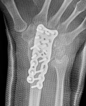
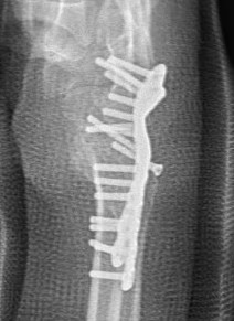
Definition
Avascular necrosis / osteonecrosis of the lunate
Epidemiology
Rare
Men 20 - 40
Anatomy
Half moon shape
- concave distally: articulates with the capitate
- convex proximally: articulates with the radius and the TFCC
Etiology
Vascular theory: repetitive micro trauma disrupting fragile vascularity of lunate
Predisposing conditions: SLE, sickle cell, Caison's
Mechanical theory: ulna minus variance causes increased radioulnar load on lunate
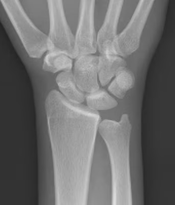
Clinical
Pain dorsal mid wrist
- stiffness
- lunate tenderness
- reduced grip strength
Xray
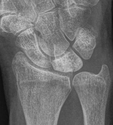
Progressive changes of AVN
- sclerosis
- fragmentation / fracture / flattenging
- midcarpal collapse: scaphoid flexion / capitate descent
- radiocarpal and midcarpal osteoarthritis
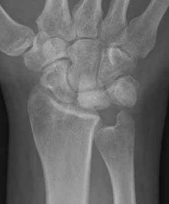
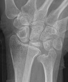
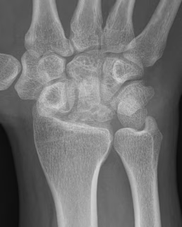
Lichtmann Classification
Stage I
- xray normal
- diagnosed on MRI
| Stage II | Stage IIIA |
|---|---|
| Sclerosis |
Collapse / fragmentation Normal carpal height |
 |
 |
| Stage IIIB | Stage IV |
|---|---|
|
Collapse / fragmentation Scaphoid flexed / Capitate migrates proximally |
Pancarpal osteoarthritis |
 |
 |
Ulna Variance
Supination and pronation alter variance
- varies up to 3 mm with wrist position
- pronation increases ulnar variance
- supination decreases variance
90 / 90 view
- zero rotation view
- neutral supination / pronation
- PA film with wrist in neutral
- elbow 90° / shoulder abducted 90°
Line from lunate fossa and ulna head
- wide variation in population
- mean ulna variance is 1 mm (range 2 - 4)
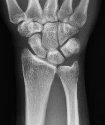
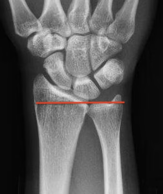
Ulna neutral
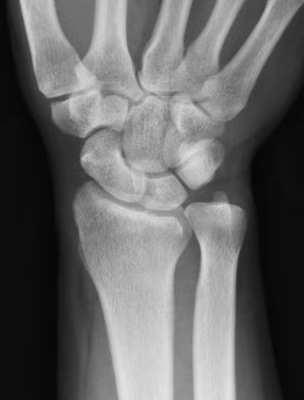
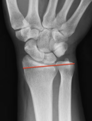
Ulna positive
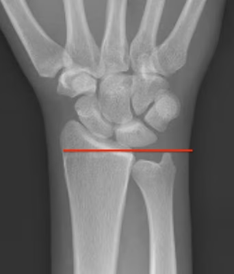
Ulna negative
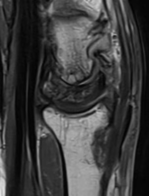
MRI
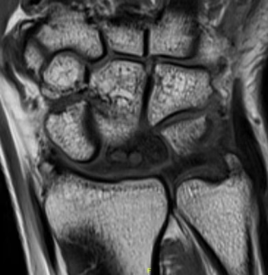
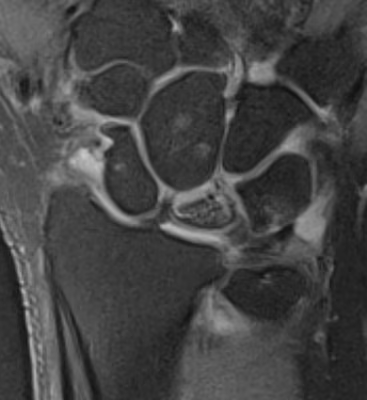
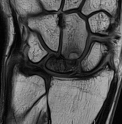
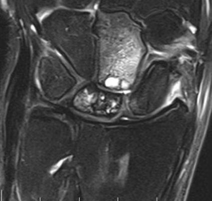
CT
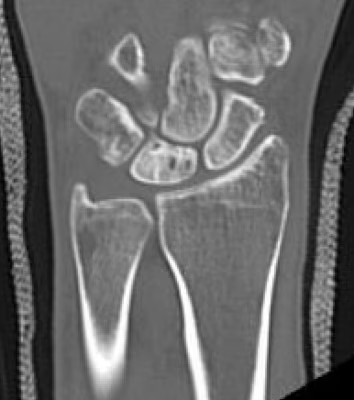
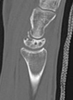
Lunate precollapse
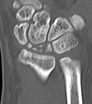
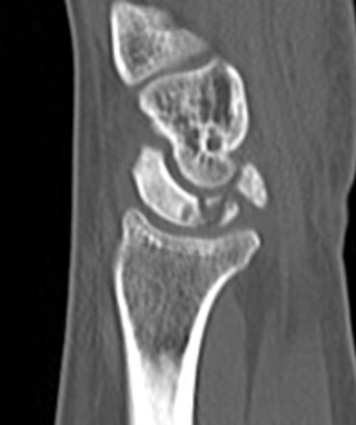
CT demonstrating lunate fragmentation and collapse
Natural history
Usually one of progressive collapse
Prognosis better in adolescents
Nonoperative management
Splint
Stage I
- trial of immobilization for 3/12 to aid revascularization
- most effective in adolescents
Operative management
Options
| Stage II / IIIA | Stage IIIB | Stage IV |
|---|---|---|
|
Radial shortening - ulna negative
Capitate shortening - ulna neutral / positive
Vascularized bone graft - ulna neutral / positive
|
Limited fusions - STT fusion - scapho-capitate fusion
Proximal row carpectomy |
Wrist arthrodesis
Wrist arthroplasty |
Stage II / IIIA
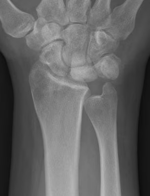
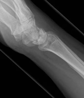
Radial Shortening
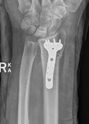
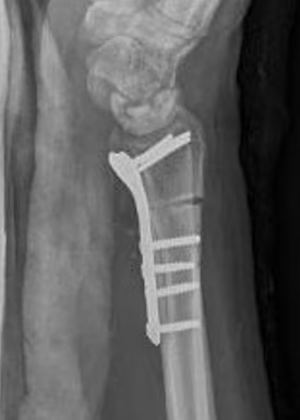
Indication
Stage II / IIIA
Negative ulna variance
Theory
Radius normally takes 80% of load
- with ulnar minus is increased to 96%
- 2mm radial shortening: 20% decrease radiolunate load
- 4mm radial shortening: 40% decrease in radiolunate load
Technique
Volar approach / bed of FCR
- osteotomy distal to DRUJ
- resection of desired amount
- aim for neutral or +1 mm ulnar variance
- cutting guides available
- volar plate
Results
- systematic review of radial shortening and nonoperative treatment
- no difference in radiographic progression
- better pain relief and ROM in radial shortening group
Quenzer et al J Hand Surg 1997
- 68 patients with radial shortening osteotomy
- reduced pain in 93%
Capitate shortening
Indication
Stage II / IIIA
Neutral or positive ulna variance
Result
Almquist Hand Lin 1993
- 83% revascularisation and healing
Vascularised Bone graft
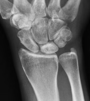
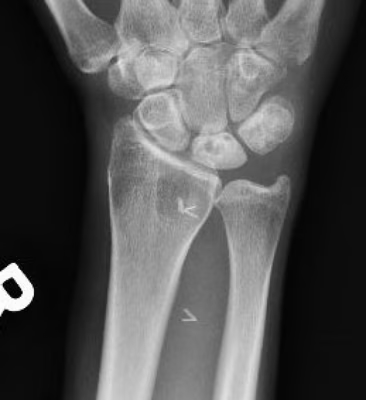
Indications
Stage II / precollapse
Ulna neutral or positive
Technique
Options
- 2nd dorsal intermetacarpal A & V
- Distal radius pronator quadratus pedicle
- Dorsal distal radius on pedicle
Results
Park et al Clin Orthop Surg 2023
- systematic review of nonoperative treatment v VBG
- reduced progression and better long term outcomes in VBG
Stage IIIB
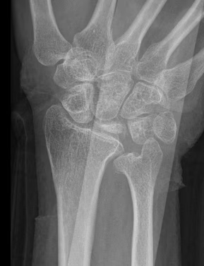
Limited fusions: STT fusion / scaphocapitate fusion
Indications
Stage IIIB with limited capitate degeneration
- reduce scaphoid flexion
- create radial column for load bearing
Technique
Dorsal approach 3/4
- resect lunate
- take scaphoid out of flexion / extend
- K wire into position
- fuse to trapezium and trapezoid or fuse to capitate
Results
Emanuels et al Plast Recon Surg 2025
- 78 scaphocapitate fusion v 64 proximal row carpectomy
- no difference in ROM or grip
- reduced pain in laborers with scaphocapitate fusion
- increased need for secondary procedures with scaphocapitate fusion
Proximal Row Carpectomy
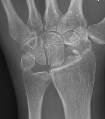
Indication
Stage IIIB
Minimal capitate degeneration
Stage IV
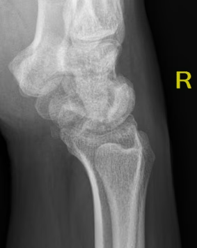
Arthrodesis
Indication
Stage IV
Stage IIIA / IIIB in manual worker