Medial Epicondyle
Ossification
Apophysis appears around 7 years
Ossifies age 16 years
Normal apophysis may be some distance from shaft
- rarely may be fragmented
Aetiology
Dislocation
Valgus deformity
Operative Indications
Absolute
- entrapped in joint
- ulnar nerve injury
- elbow dislocation
Controversy
Displacement > 5mm
- good outcomes reported up to 10 mm
- clinically have some valgus instability
- non significant to most patients
- may be significant in the throwing athlete
Farsetti JBJS Am 2001
- 42 patients with displacment > 5 mm
- average age 12 years
- half treated in long arm cast, half ORIF
- no difference in outcome
- poor outcome with fragment excision and ligament reattachment
- in nonoperative group all but two had nonunion radiographically
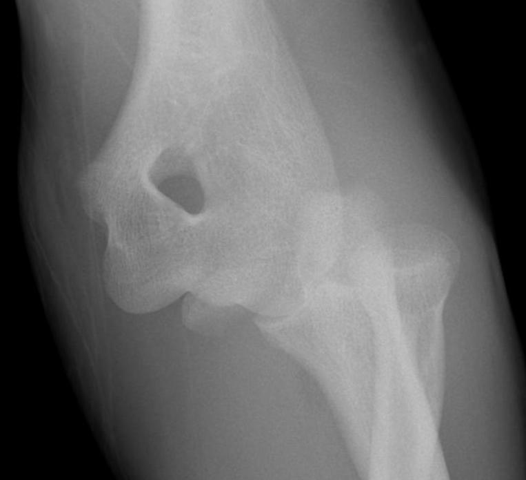
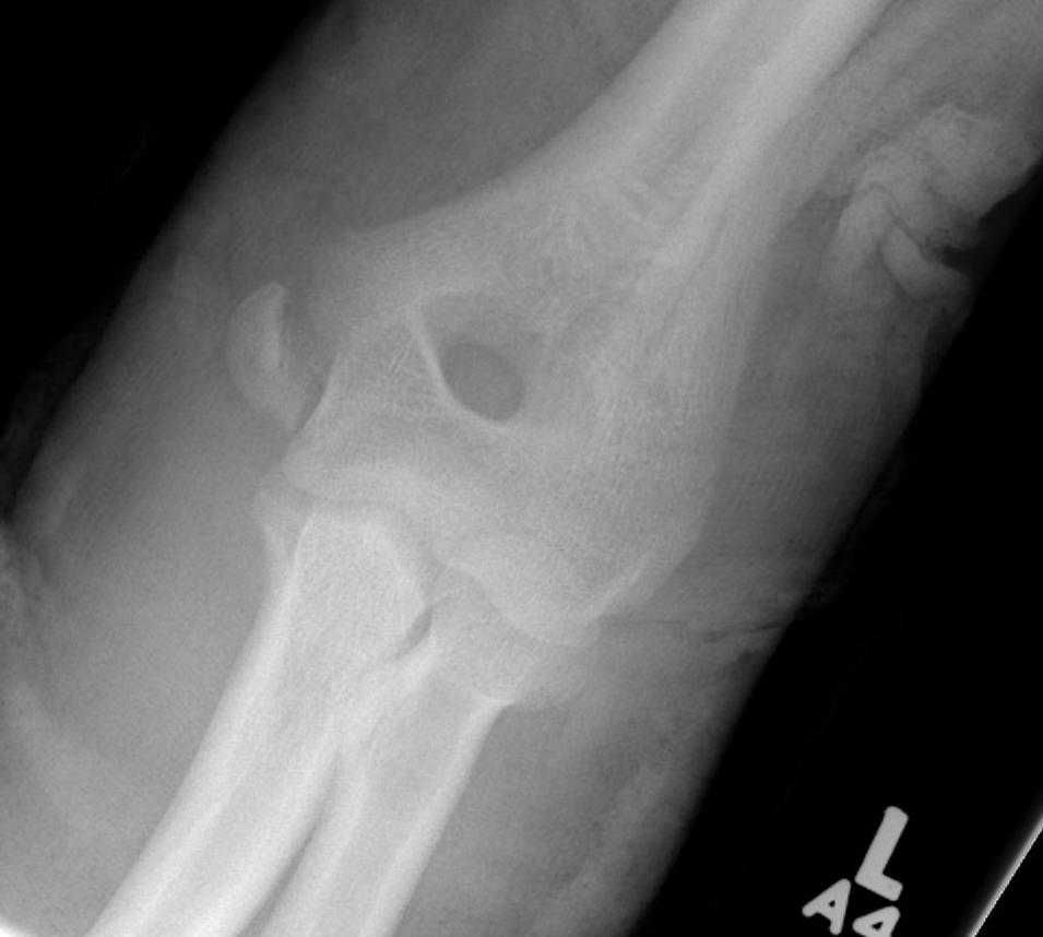
Entrapped in joint
Aetiology
- secondary to dislocation which has self reduced
Issue
< 5 with dislocation
- medial epicondyle not visible
- may be entrapped in joint
- suspect if decreased ROM / pain / non congruent reduction
- can try arthrogram
- may nee open exploration of joint
Management
Can attempt MUA
- valgus and extend wrist to tighten flexor origin
- rarely works
Usually require open reduction + K wire
ORIF
Technique
Position
- usually supine with arm table
- can place patient prone which makes fragment very easy to reduce
Medial incision
- find and protect ulna nerve
Reduce fragment anatomically
- ORIF with K wires or screw
Medial Condyle Fracture
Epidemiology
Rare
- can be difficult to diagnose
- trochlea may not be ossified
- looks like medial epicondyle injury
Management
ORIF if displaced > 2 mm
- intra-articular fracture
- may require MRI
Complications
Nonunion / cubitus varus
Trochlea AVN
Medial overgrowth / cubitus valgus