closed reduction
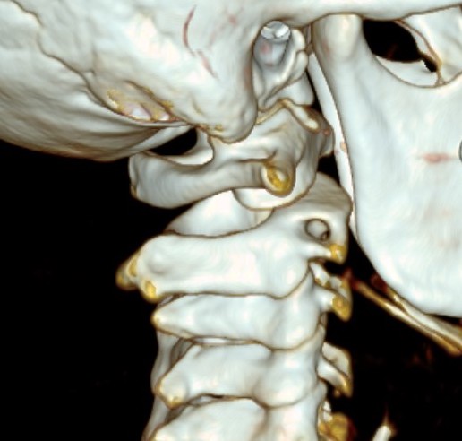
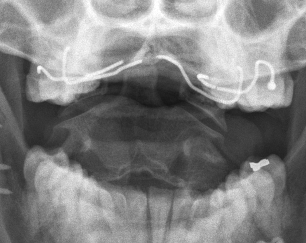
Distal humeral physeal separation
Pathology
Children < 6
- entire distal humerus physis is displaced
Xray
Distal physis not ossified < 1 year
- may be a difficult diagnosis
Management 6 - 18 months
Two groups of dislocated hips
1. Late presenters
2. Failures of splint in those < age 6/12
Options
1. Adductor tenotomy + closed reduction
- most surgeons will attempt this initially
- risk of AVN wilth forceful reduction / excessive abduction
2. Open Reduction
- for failure of closed reduction
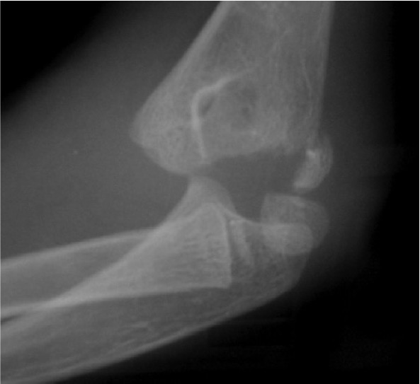
Radial neck fracture
Mechanism
FOOSH
- valgus injury
- don't get radial head fracture as is mostly cartilaginous
Types
SH 1 or 2
Associated Injuries
MCL injury
Olecranon / Medial epicondyle fracture
Management
Deformity
Varus / extension / external rotation
Options
Intertrochanteric
Base of Neck
Subcapital
Osteotomy
Valgus / flexion / internal rotation
Intertrochanteric / Southwick
Technique
- biplanar
- valgising / flexion / internal rotation
Background
Definition
Displacement of proximal femoral epiphysis in the immature hip
- due to imbalance of mechanical and endocrine factors
Epidemiology
Age Peak Incidence : M 12-14; F 11-13; Slight downward trend due to earlier maturation of children
L hip > R
10 / 100 000
Bilateral SUFE
No endocrine abnormality
- 20% at time of of diagnosis
- another 20% during diagnosis
- up to 60% with long term follow up
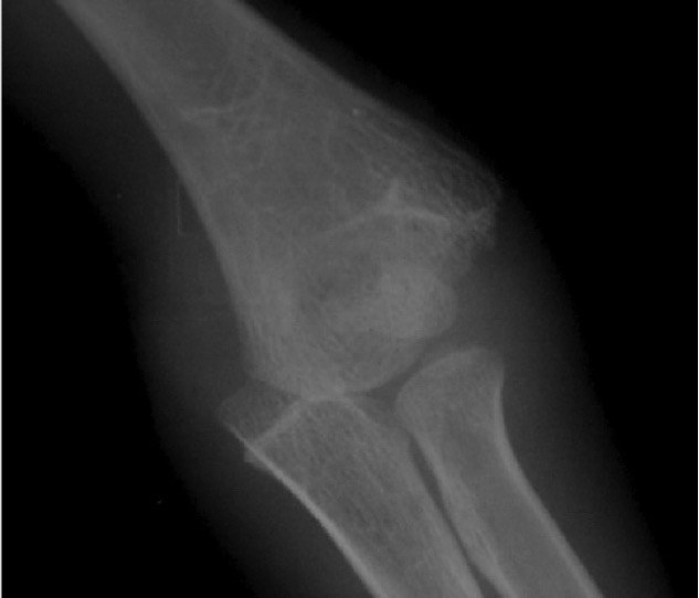
Lateral condyle fractures
Epidemiology
Average age 6 years
20% distal humeral fracture
- second most common elbow fracture after supracondylar
Mechanism
Pull Off
- more common
- fracture begins posterolateral metaphysis
- LCL, ECRL & ECRB attached to fragment
Push off
- varus force to extended EJ
Classification