Definition
Angular deformities / coronal plane deformities
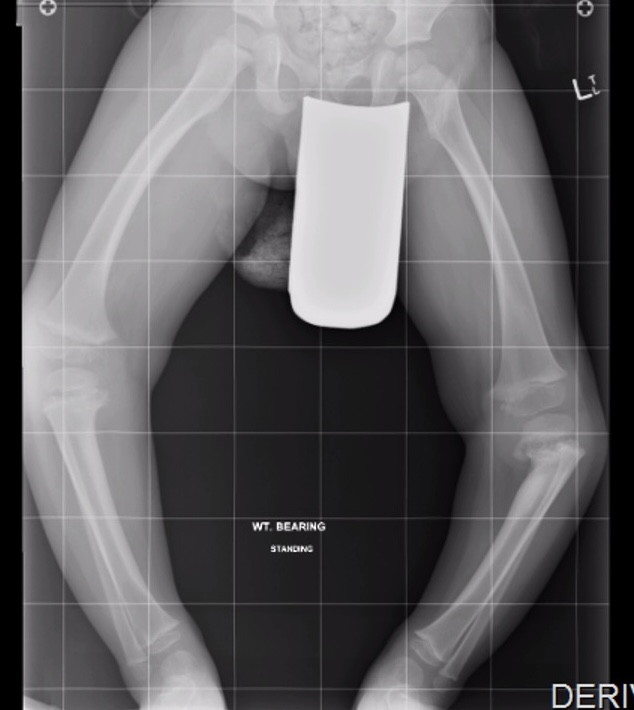
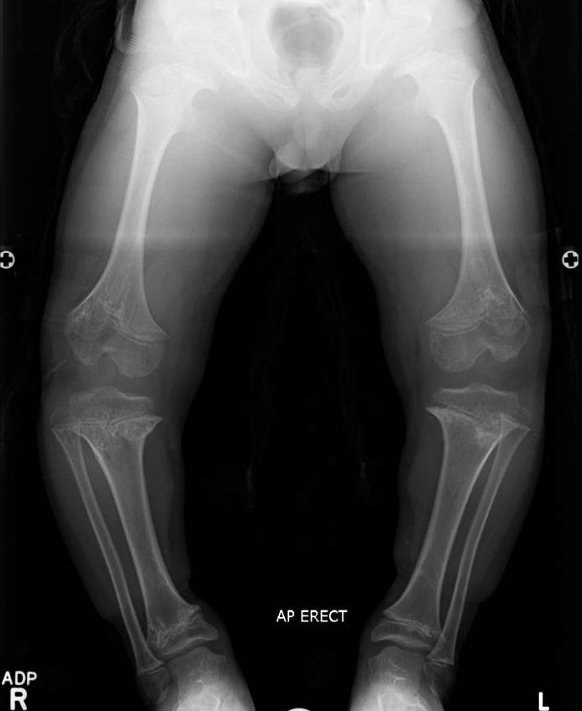
- varus / bow legs
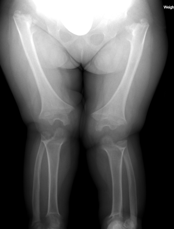
- valgus / knock knees
Causes
| Physiological | Pathological varus | Pathological valgus |
|---|---|---|
| Normal development |
Blount's Rickets
Skeletal dysplasia Anterolateral bowing - pseudoarthrosis Fibrous dysplasia Tibial hemimelia |
Congenital posteromedial bowing Anteromedial bowing / fibular hemimelia Cerebral palsy / spina bifida Lateral condylar hypoplasia |
| Within Salenius curve | Rickets | Rickets |
| Growth plate damage - trauma / infection | Growth plate damage - trauma / infection | |
| Juvenile rheumatoid arthritis | Juvenile rheumatoid arthritis | |
| Osteogenesis imperfecta | Osteogenesis imperfecta | |
| Skeletal dysplasia | Skeletal dyplasia | |
| Multiple osteochondromatosis |
Issues
Excessive valgus - patellofemoral instability
Excessive varus - medial osteoarthritis
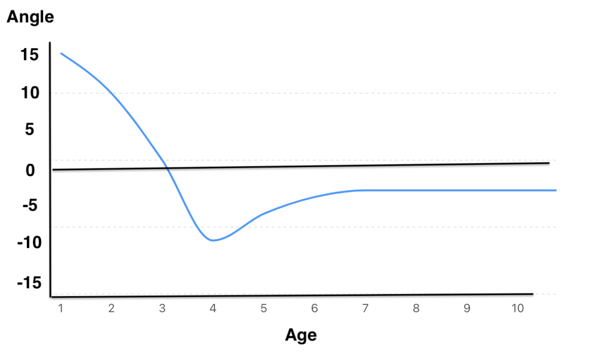
Normal development
Salenius and Vankka et al JBJS Am 1975
- tibiofemoral angle in 1400 children over time
- demonstrated that there is a standard progression over time
- varus to neutural to excessive valgus to physiological valgus
Birth to 18 months: physiological varus / bow legs
18 - 24 months: straight legs
2 - 4 years: knock knees / 10° valgus
4 - 6 year: physiological valgus / 6° valgus

3 years 6 years
Salenius curve
- range ~ 15° either way at each age
- map child against this curve over time
- ensure normal progression / variation
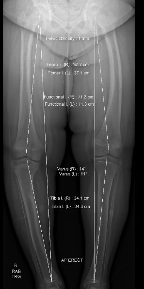
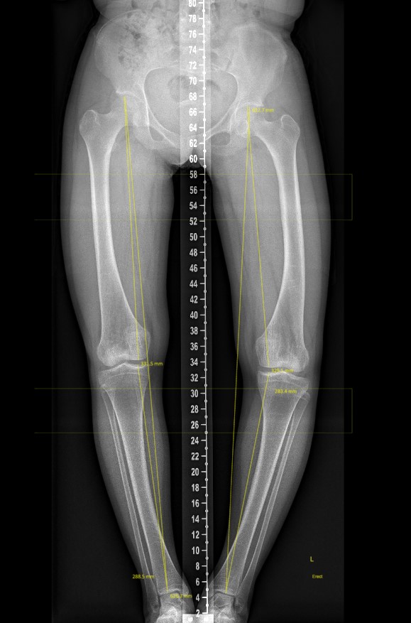
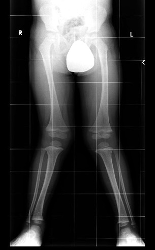
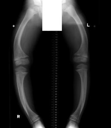
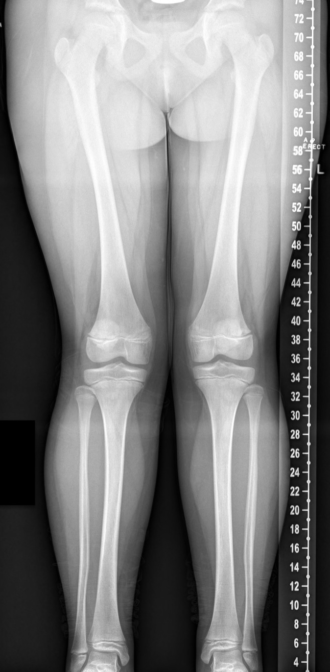
Examination
Standing
- unilateral / bilateral
- varus / neutral / valgus
- inter-condylar distance
Leg length
Rotational profile
- inter-malleolar angle - external tibial torsion
- proximal femoral anteversion - hip internal rotation
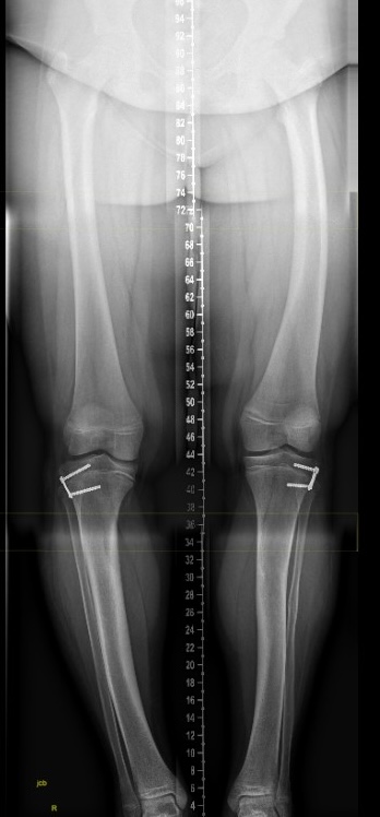
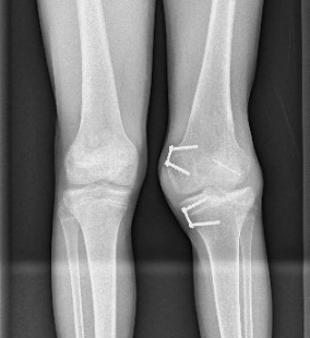
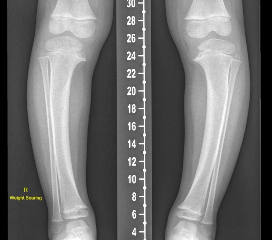
X-ray
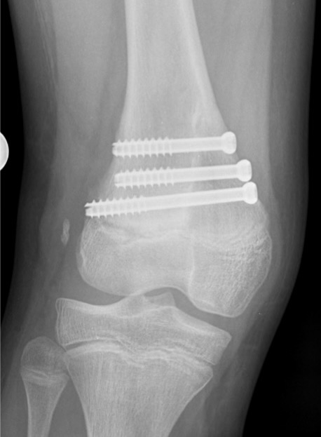
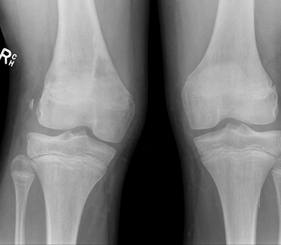
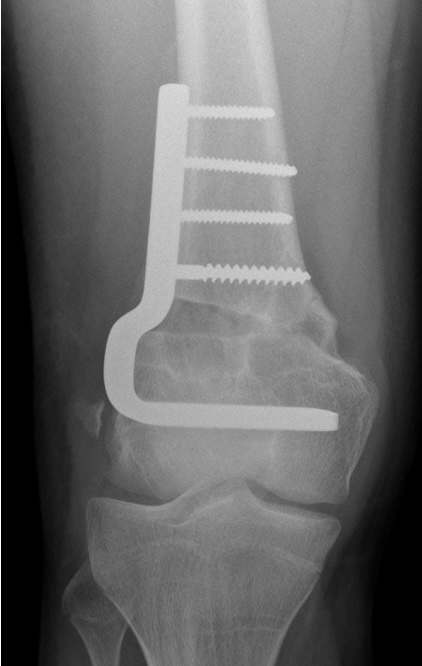
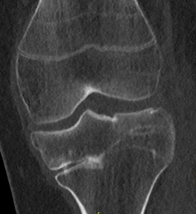
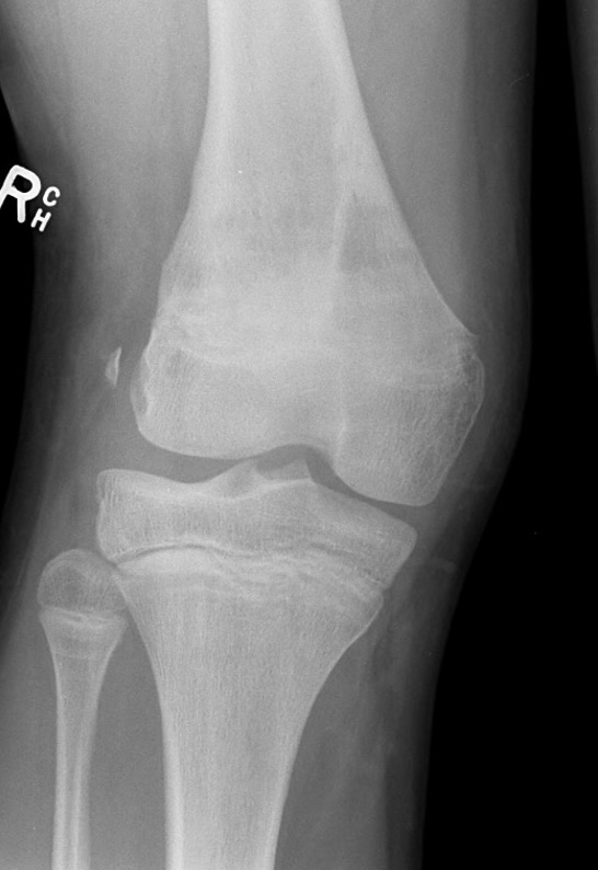
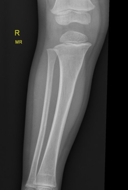
Growth plate injury / trauma
| Proximal tibial fracture | Distal femur fracture | Couzen's proximal tibia fracture |
|---|---|---|
 |
 |
 |
Rickets
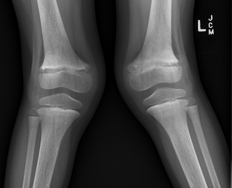
Blounts
Other
| Multiple osteochondromas | Achondroplasia | Multiple epiphyseal dysplasia |
|---|---|---|
 |
 |
 |
Management
Options
Partial growth plate arrest
- physeal bar resection
- > 2 years growth and < 50% physeal bar
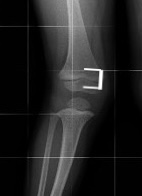
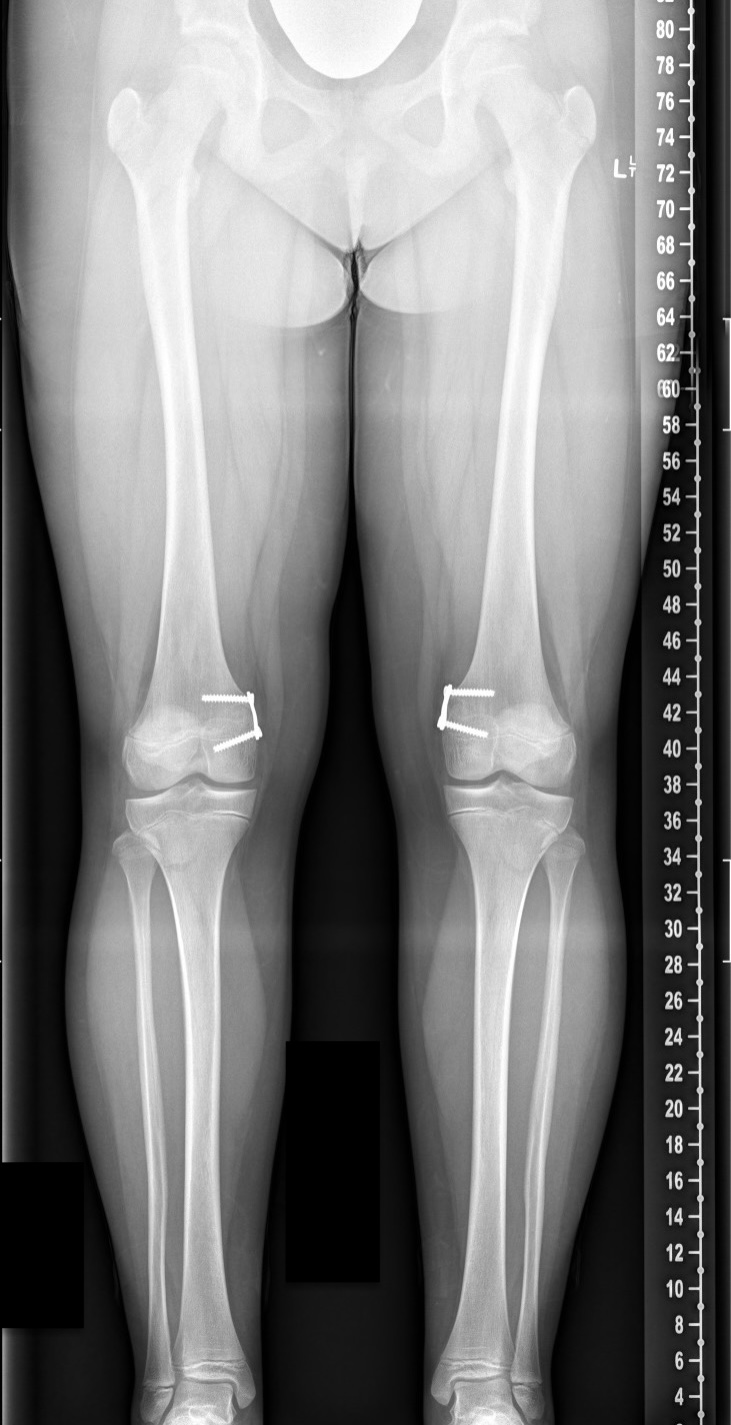
Angular deformity
- guided growth / growth modulation
- hemi-epiphysiodesis
- osteotomy - skeletally mature
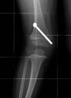
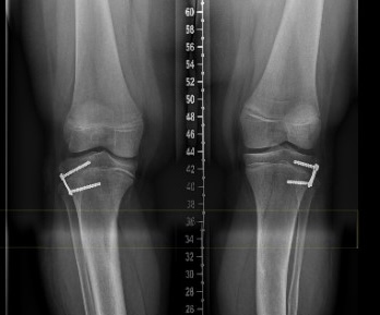
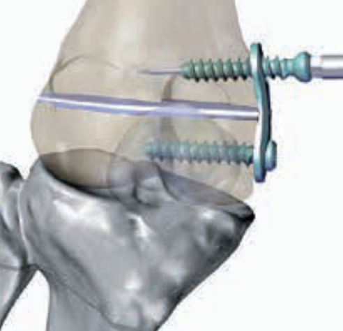
Guided growth / growth modulation
Concept
Temporary growth cessation
- tension band plating of epiphysis / 8 plates
- transphyseal screw
Technique
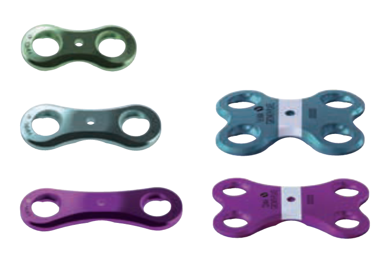
Eight plate surgical technique PDF
Arthroscopy techniques guided growth PDF
Results
Tension band plating
- systematic review of Eight plates for coronal plane deformity
- 7 studies and 350 limbs
- mean age 10
- 33% idiopathic, 67% pathological
- successful correction 91%
- deformity correction 1.3 °/month
- lateral distal femoral angle 0.9°/month
- medial proximal tibial angle 0.7°/month
- complications 6%: hardware failure, overcorrection, stiffness - rebound 4%
Screw versus tension band plating
Sabry et al BMC Musculoskeletal Disord 2025
- systematic review of tension band plating v percutaneous transphyseal screw
- correction of coronal plane deformity
- screw faster correction and lower complications
Rebound
Eberle et al J Child Orthop 2023
- 189 legs with idiopathic genu valgum treated with tension band plating
- 59% rebound growth
- 30% stable
- 11% continuous correction
Osteotomy