
Trochanteric osteotomy
Indication
Increases exposure to acetabulum
Assists with removal femoral implants and cement
Types
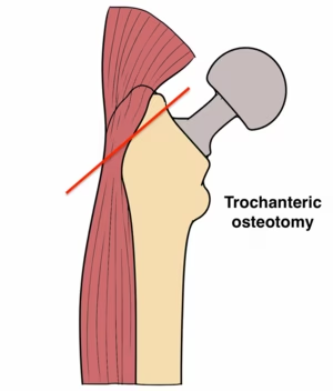
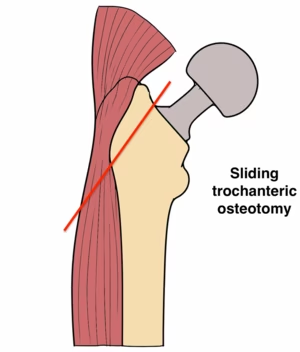
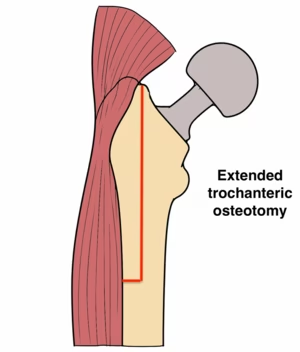
| Trochanteric osteotomy | Sliding trochanteric osteotomy | Extended trochanteric osteotomy |
|---|---|---|
|
Osteotomy greater trochanter Only abductors attached |
Osteotomy greater trochanter Abductors and vastus lateralis attached |
Osteotomy of greater trochanter and 10 - 15 cm of femur |
|
Inherently unstable High risk nonunion |
Increased stability |
Indicated for removal of femoral implants and cement Must bypass osteotomy with long femoral implant |
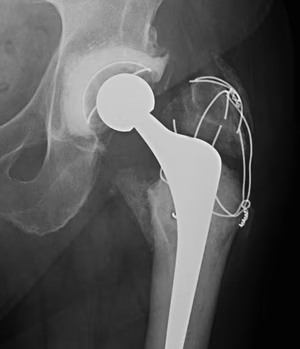
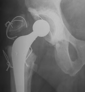
Standard trochanteric osteotomy
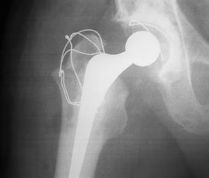
Background
Detach greater trochanter with only abductors attached
- difficult to repair as more vertically unstable with lateralis released
Risk of nonunion - risk of painful hardware / limp / dislocation
Technique
Detach proximal attachment of vastus lateralis
- pass retractor deep to abductors and superficial to capsule
- osteotomy from lateral aspect of GT angled up towards pirifiromis fossa
- consider biplanar / Chevron osteotomy to increase union rates
- detach any short external rotators and reflect superiorly
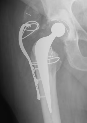
Fixation - wires / cables / grip plate
Results
- flat osteotomy versus Chevron osteotomy
- 69 flat osteotomy: 11% nonunion
- 69 biplane osteotomy: 2% nonunion
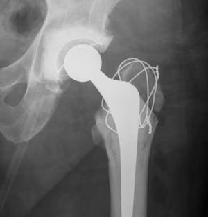
Trochanteric slide
Background
Vastus lateralis and abductors left attached to fragment
- fragment retracted anteriorly
- increased inherent stability
- vastus lateralis prevents proximal migration
Technique
Retractor superiorly deep to gluteus minimus
Retractor deep to insertion of vastus lateralis
Osteotomy anterior to posterior
Fixation - wires / cables / grip plate
Results
- trochanteric slide in 94 revision THA
- 96% union rate
Extended trochanteric osteotomy (ETO)
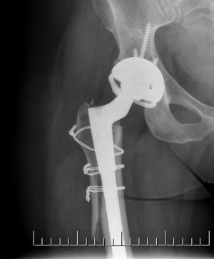
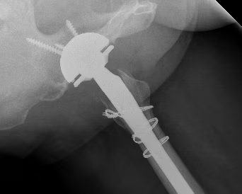
Background
Osteotomy greater trochanter and anterior proximal femur
- posterior to anterior longitudinal cut
- 10 - 15 cm long and 1/3 to 1/2 diameter of femur
- short distal transverse cut
- hinges open anteriorly
Technique
Measure length from greater trochanter
- elevate vas lateralis /abductors to expose linea aspera / distal femur
- osteotomy posterior cortex
- transverse cut 1/3 diameter
- osteotomy or drill holes anterior
- lever osteotomy open anteriorly
Fixation
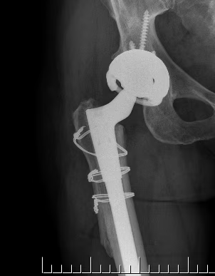
- 3 x cerclage cables submuscular
- protect sciatic nerve
Results
Malahias et al J Arthroplasty 2020
- systematic review of 1500 ETO
- union rate 93%
- subsidence rate > 5 mm 7%
Implant removal
Cemented femoral implant
Stem often easily removed if cemented
- extraction devices hook around proximal prosthesis
- can release cement – implant interface with flexible osteotomes / micro sagittal saw / small burr
Cement removal
- may require extended trochanteric osteotomy
- cement removal kits with flexible osteotomes, reverse hooks, cement splitters
- split cement radially and then remove
- remove cement plug
Uncemented femoral implant
Can be very difficult to remove a well fixed stem in setting of infection
- femoral implant may be proximally coated or extensively coated
- breakdown osseointegration with flexible osteotomes and microsagittal saw
- may require extended trochanteric osteotomy
Uncemented acetabulum
AO Foundation acetabulum removal
Liner
Option 1. Company specific removal instruments / disengage locking mechanism
Option 2. Lever out liner with osteotomes
Option 3. Drill hole in liner with 4.5 mm drill, insert 6.5 mm screw to push liner out
Metal Shell
Curved osteotomes
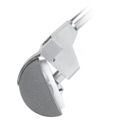
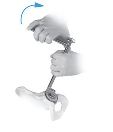
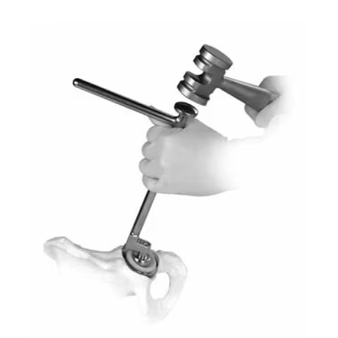
Zimmer Explant Acetabular Removal System
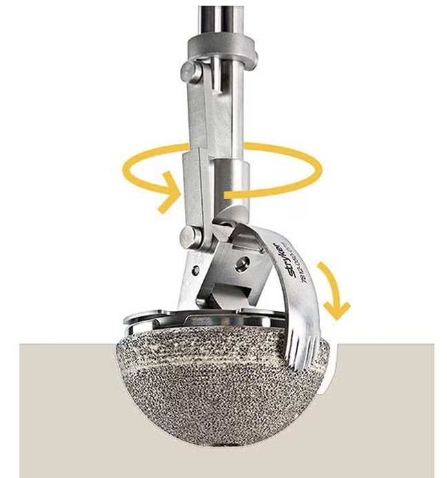
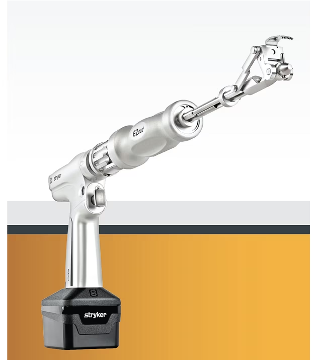
Styker EZout Powered Acetabular Revision System
Technique
- must remove screws first, then replace liner
- central head sized to sit in liner
- diamond blades cut between cup and bone
- initial blade short
- second is thin and full radius
Cemented acetabulum
Option 1. Loosen poly cup from cement with curved osteotomes
Option 2. Ream out the poly cup
Option 3. Insert threaded extractor through drill hole in poly and remove