
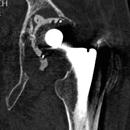
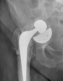
Indications for revision
Loosening
Infection
Instability
Periprosthetic fracture
Pain
Preoperative Assessment
Exclude infection
Quantify bone loss
Femoral Bone Loss
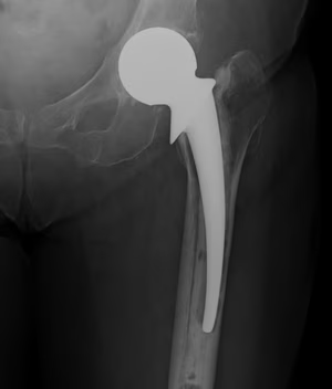
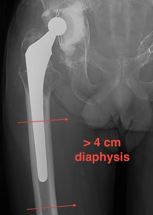
Paprosky classification
| Type I |
Minimal metaphyseal cancellous bone loss Intact diaphysis |
|---|---|
| Type II |
Extensive metaphyseal cancellous bone loss Intact diaphysis |
| Type IIIA |
Metaphysis severely damaged > 4cm diaphyseal bone for distal fixation |
| Type IIIB |
Metaphysis severely damaged < 4cm diaphyseal bone for distal fixation |
| Type IV |
Extensive metaphyseal and diaphyseal bone loss Isthmus non supportive |
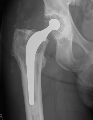
Paprosky Type I
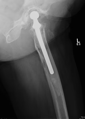
Paprosky Type II
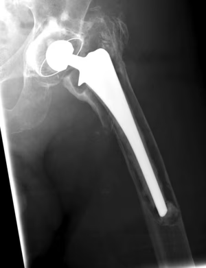
Paprosky Type IV
Acetabular bone loss
4 radiographic criteria
Superior migration of the hip center
Ischial osteolysis - posterior column damage
Teardrop osteolysis and medial migration - medial wall damage
Position of the implant relative to Kohler’s line - deficiency of anterior column
Paprosky classification
Paprosky et al J Arthroplasty 1994
Based on severity of bone loss and ability to obtain cementless fixation with intact rim
- Type I: minimal deformity with intact rim
- Type II: > 50% bone to support cup
- Type III: Non supportive rim
|
Femoral head center migration |
Ischial osteolysis |
Kohler line | Teardrop | ||
|---|---|---|---|---|---|
| Type I | None | Intact | Intact | Intact |
Minimal deformity with intact rim Medial wall / anterior & posterior column intact No superior migration hip center |
| Type IIA |
Mild < 3cm |
None | Intact | Intact |
Superior bone lysis Superior rim intact |
| Type IIB |
Moderate < 3cm |
Mild | Intact | Intact |
Superior rim deficient Superolateral migration |
| Type IIC |
Mild < 3 cm |
Mild | Disrupted | Moderate | Medial defect but wall intact |
| Type IIIA |
Severe > 3cm |
Moderate | Intact |
Moderate lysis |
Severe > 2 cm superolateral migration Medial wall and posterior column lysis Bone loss 10 - 2 o'clock |
| Type IIIB |
Severe > 3cm |
Severe | Disrupted | Severe |
Severe > 2 cm superomedial migration Damage to medial wall / posterior & anterior column Bone loss 9 - 5 o'clock |
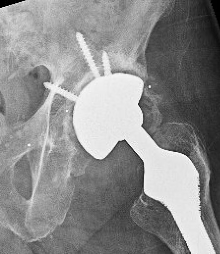
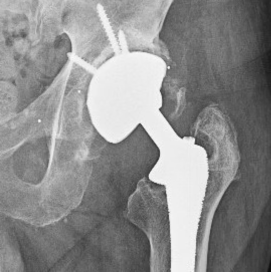
Paprosky Type I: Minimal bone loss with intact rim
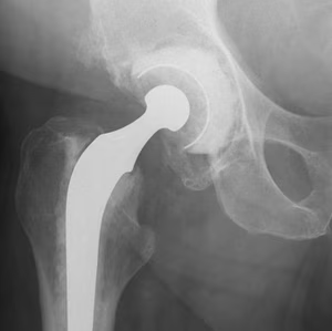
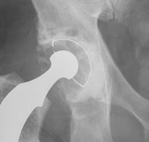
Paprosky Type IIB: Superior rim deficient with superolateral migration
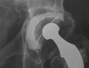
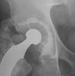
Paprosky Type IIC: Medial lysis and migration but medial wall intact
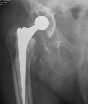
Paprosky Type IIIA: Superolateral migration
AAOS Classification
| Type I |
Segmental deficiencies - peripheral: superior / anterior / posterior - central: medial wall |
|---|---|
| Type II |
Cavitary deficiencies - peripheral: superior / anterior / posterior - central: medial wall |
| Type III | Combined deficiencies |
| Type IV |
Pelvic discontinuity - separation anterior & posterior columns |
| Type V | Arthrodesis |
Pelvis discontinuity
Xray signs
- fracture line seen on two xray views (AP and lateral)
- single fracture line with medial pelvic translation
- medial migration implant
CT
- can miss discontinuity due to metal artefact
- oblique reconstructions increase sensitivity from 70% to 90%
CT angiogram
- intra-pelvic component / cement
- beware catastrophic vascular damage with component removal
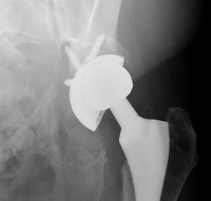
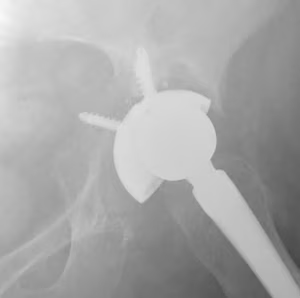
Fracture line seen on two xray views
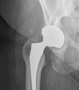
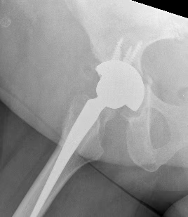
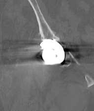
Fracture line confirmed on CT
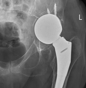
Intrapelvic migration of components