Complications
Complications
Types
Chondrolysis
AVN

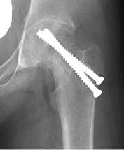
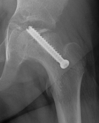
Chondrolysis
AVN


Much less common than hip and knee OA
Patients tend to be younger
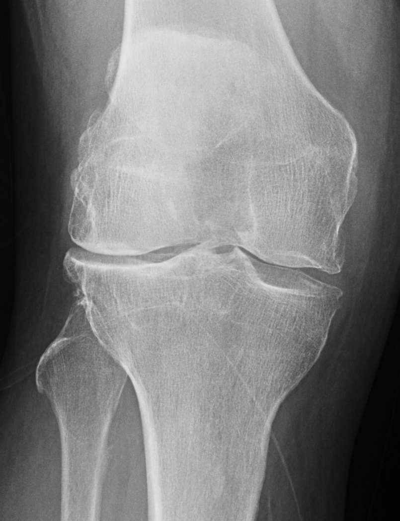
Trauma
RA
Rickets / osteomalacia
Horizontal joint line important
- < 10° tilt acceptable
> 10o joint line tilt / due to femoral valgus
- continues to overload lateral compartment
Stage 0
Natural history mixed
- depends on size of lesion and diagnosis
- treat if becomes asymptomatic
- may benefit from bisphosphonates
Stage 1 / Normal X-ray, abnormal MRI
Forage: 80% G/E
Bisphosphonates
Stage 2 / Abnormal X-ray with cysts and sclerosis
A: As for Stage I
Metabolic Bone Disease
Paget's
Fracture Malunion
Previous Osteotomy
1. Intra-articular correction
2. Simultaneous osteotomy and TKR
3. Staged correction and TKR
Femur
- draw line of mechanical axis of femur
Hip has been out for some time
- degree of acetabular dysplasia evident
- less time for remodelling
- increased instability if not addressed
Open reduction + FDRO / Pelvic Osteotomy
- usually perform pelvic osteotomy to correct acetabular dysplasia
- reserve FDRO for > 3 years / or if difficult reducing hip

Progressive varus deformity of knees
- secondary to abnormality of medial upper tibial physis
- localised varus & internal rotation deformity
Infantile form
- onset 1-3 years / bilateral
Adolescent form