Cozen fracture
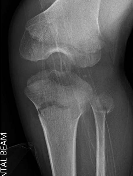
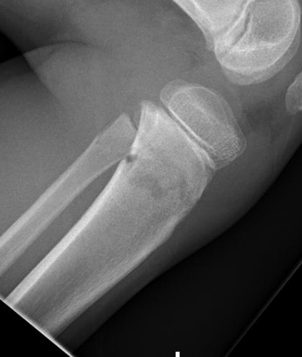
Definition
Proximal metaphyseal tibial fracture
Risk of delayed valgus deformity

Proximal metaphyseal tibial fracture
Risk of delayed valgus deformity
Theory
- want to traverse one muscle / one compartment
- keep away from NV bundle
- as a rule perform open biopsy through compartment the tumour is in
- this is the compartment that will require surgical removal in wide excision
- direct approach without going through muscle if possible i.e. tibia, distal ulna
Thigh
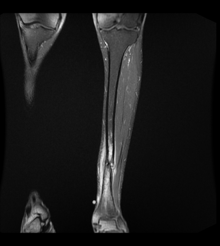
Athletic / high impact exercises
First described in ballet dancers (Burrows 1956)
- tension side of bone / lateral side
- progression to complete fracture has been well documented in athletes
Point tenderness
- lateral aspect of tibia
Over time develop bony lump
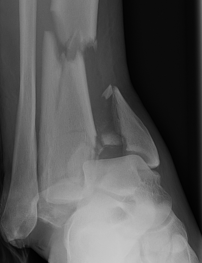
Complex / high energy injuries
Management of soft tissues critical
- restore length with external fixation
- await for swelling to reduce
Restoration of alignment & joint surface imperative
Outcome guarded
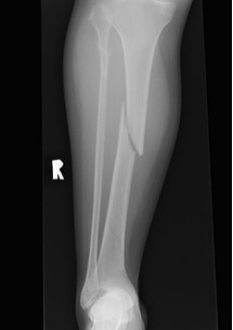
Most common long bone fracture
Young patients / sports
Elderly / simple falls
MVA - often compound
Grade 0
- nil ST injury
Grade 1
Acceptable reduction
- varus / valgus < 5o
- anterior / posterior < 5o
- rotation 5o
- shortening 10 mm
Poor remodelling potential
- valgus
- apex posterior angulation / recurvatum
- rotational alignment does not remodel
- shortening / in 2-10 year old average overgrowth is only 5mm
Anterolateral bowing
True bone dysplasia
- hamartomatous area in the tibia
- non union or potential non union through pathological fracture in this region
The defect in the bone is filled with mature, fibrous connective tissue
- not really a pseudoarthrosis
- by definition, a pseudarthrosis has a cleft lined by fibrocartilage containing fluid and bounded by a capsule