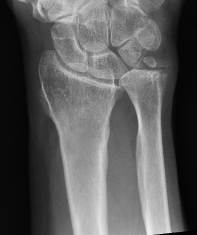
Definition
Unacceptable position of distal radius post fracture causing pain / stiffness / loss of function
Types
- extra-articular
- intra-articular
- mixed
Incidence
Raudasoja et al J Hand Surg Eur 2024
- national database of 41,000 distal radius fractures
- 300 oseotomies
- highest in those aged 40 - 50
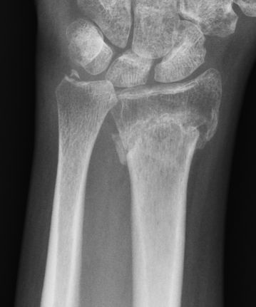
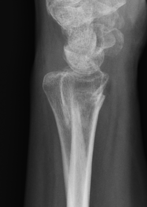
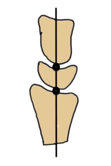
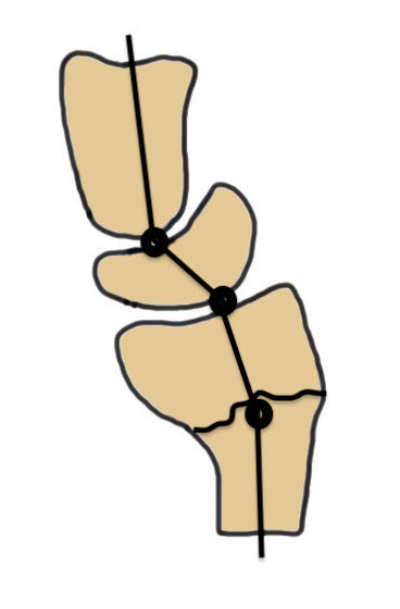
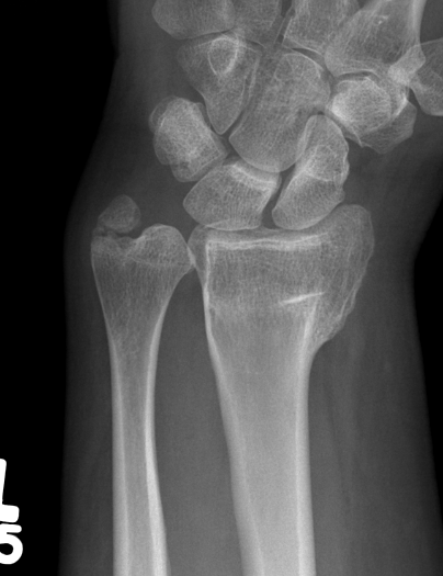
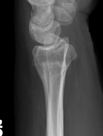
Malunion
| Radial shortening | Radial inclination | Positive ulna variance |
|---|---|---|
|
|
|
|
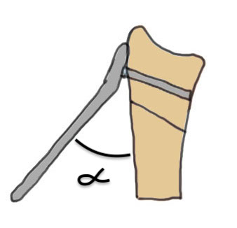
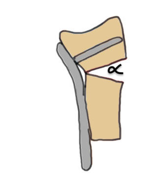
| Dorsal tilt > 15 degrees | Volar tilt > 10 degrees | Articular step > 3 mm |
|---|---|---|
|
|
 |
 |
Pathology
Radial shortening
- affects normal kinematics of the DRUJ
- ulnocarpal abutment
Dorsal tilt
- loss of flexion
- +/- midcarpal instability - DISI / CIND without interosseous ligament disruption
Clinical
Stiffness - loss of dorsiflexion / supination
Weak grip
Ulna sided pain
Wrist pain
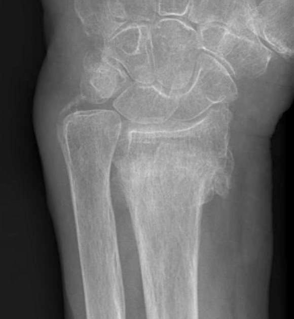
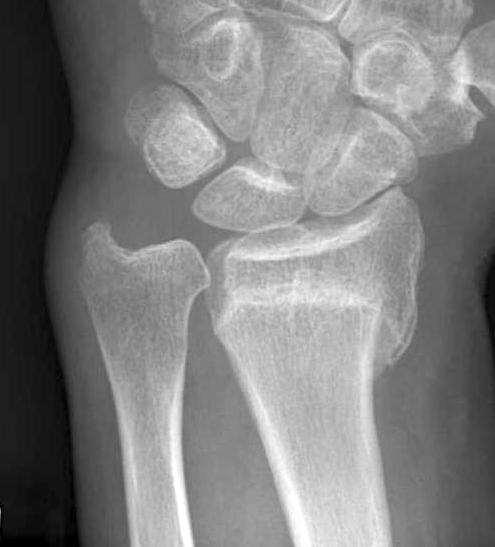
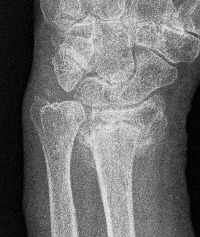
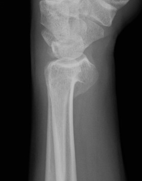
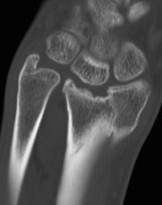
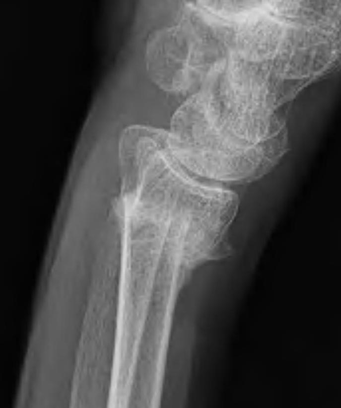
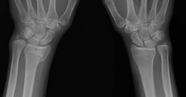
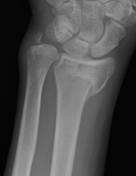
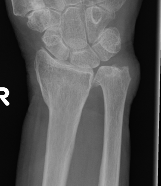
Xray
Bilateral xrays
PA film in neutral
- wrist neutral
- elbow & shoulder at 90°
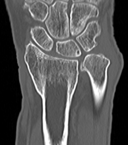
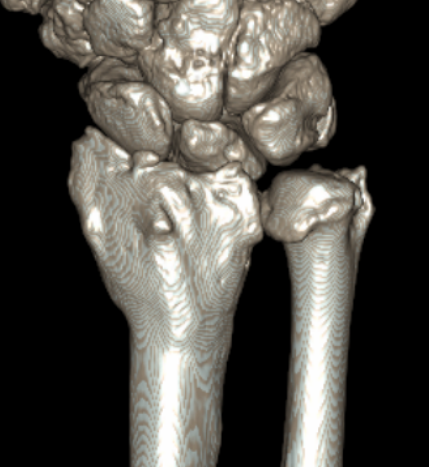
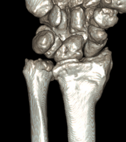
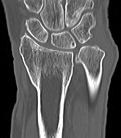
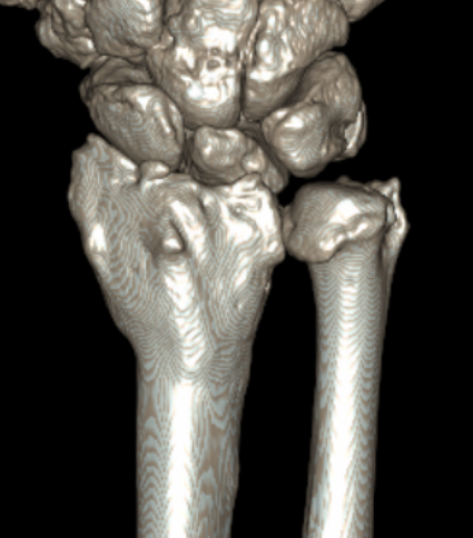
CT
Operative Management
Indications
Pain
Disability
Contraindications
Radiocarpal osteoarthritis
Osteoporosis / smoking
CRPS
Surgical Options
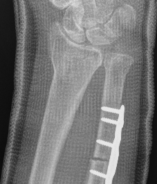
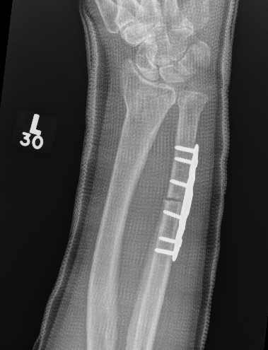
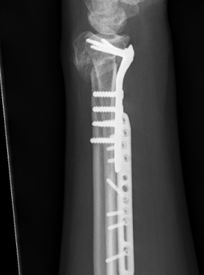
Ulna shortening
Radial osteotomy - volar versus dorsal
Radial osteotomy + ulna shortening +/- distal ulna resection / fusion
Intra-articular osteotomy
Soft tissue releases for stiffness
Outcomes
Radial osteotomy versus ulna shortening
Ma et al Arch Orthop Trauma 2022
- 68 patients with radial malunion
- radial lengthening versus ulna shortening
- better pain relief and functional outcomes with radial osteotomy
- shorter surgery with ulna shortening
- 2 revisions for painful nonunion with ulna shortening
Radial osteotomy versus DRUJ fusion
Wang et al J Hand Surg Am 2024
- RCT of 33 patients > 60 years old with radial malunion
- radial osteotomy versus Suave-Kapanji
- better grip strength in radial osteotomy
- similar outcomes in both groups
- shorter operative times with Suave-Kapanji
Ulna shortening
Low et al Arch Orthop Trauma 2014
- 23 patients 7 years post ulna shortening osteotomy
- 21/23 satisfied
- better results with minor radial displacement (< 10 degrees)
- better results with postoperative ulna positive or neutral
Volar versus dorsal radial osteotomy
- 28 patients undergoing osteotomy for radial malunion
- dorsal plate: increased complications including plate removal
- volar plate: increased undercorrection, difficulty with plate fitting
CT guided 3D planning
- RCT of 40 patients with radial malunion
- 2D versus 3D planning and patient specific surgical guides
- 3 degree better correction in 3D group
- non significant trend towards better outcomes in 3D group
Ulnar Shortening
Indications
Short radius, positive ulna variance
Acceptable alignment distal radius
Acceptable DRUJ
Technique
Approach to ulna
- between ECU and FCU
- can use cutting jigs
- resect 2 - 6 mm of ulna based on xray templating
- compression plate
Vumedi ulna shortening osteotomy using cutting jig video
Medartis ulna shortening jig PDF
Acumed ulna shortening jig PDF
Results
Owens et al J Hand Surg Am 2019
- systematic review of ulna shortening osteotomy
- nonunion rate 4%
- no difference between transverse or oblique osteotomy
- delayed union: transverse 7%, oblique 4%
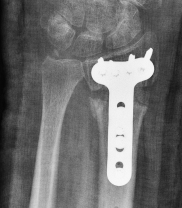
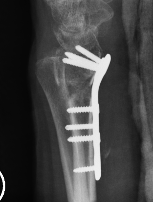
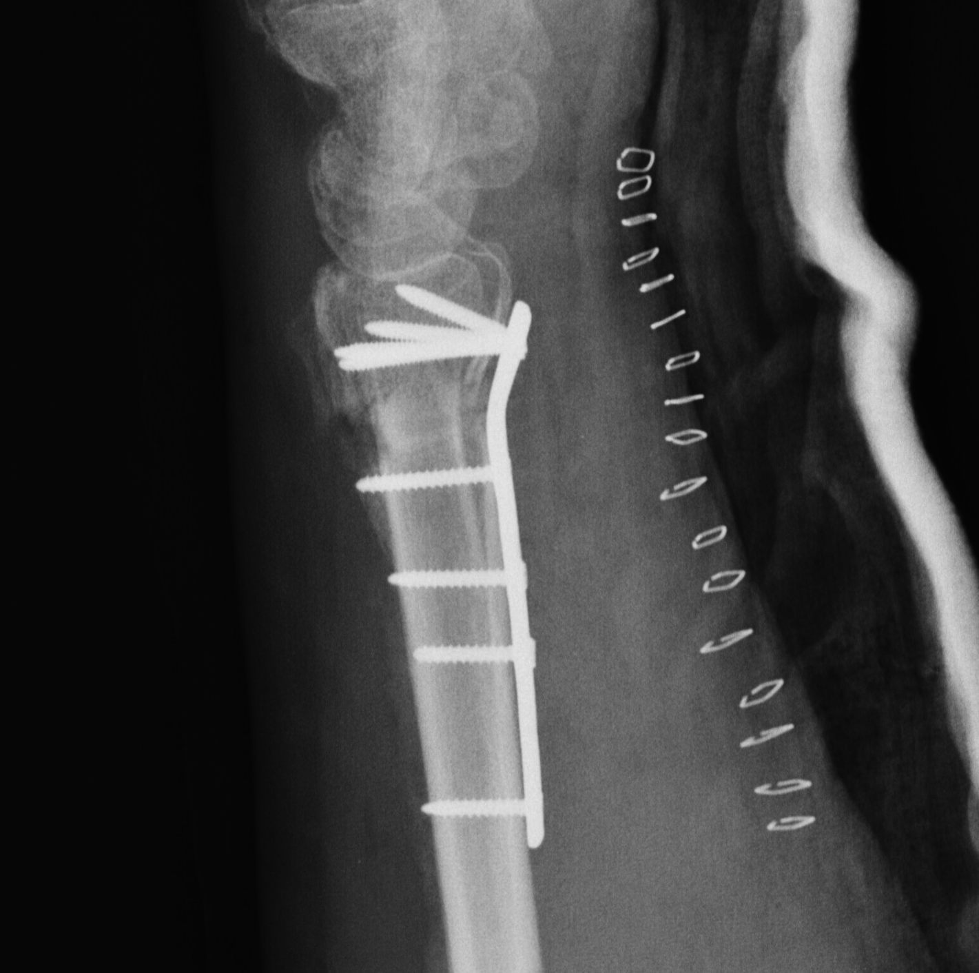
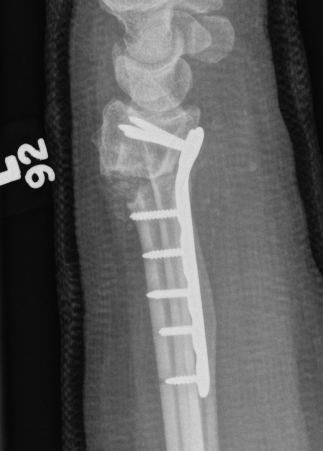
Distal radial osteotomy
Indications
Dorsal tilt / radial tilt / loss of inclination
Acceptable DRUJ articular surface
Options
Dorsal opening wedge
Volar opening / closing wedge
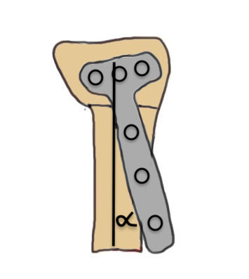
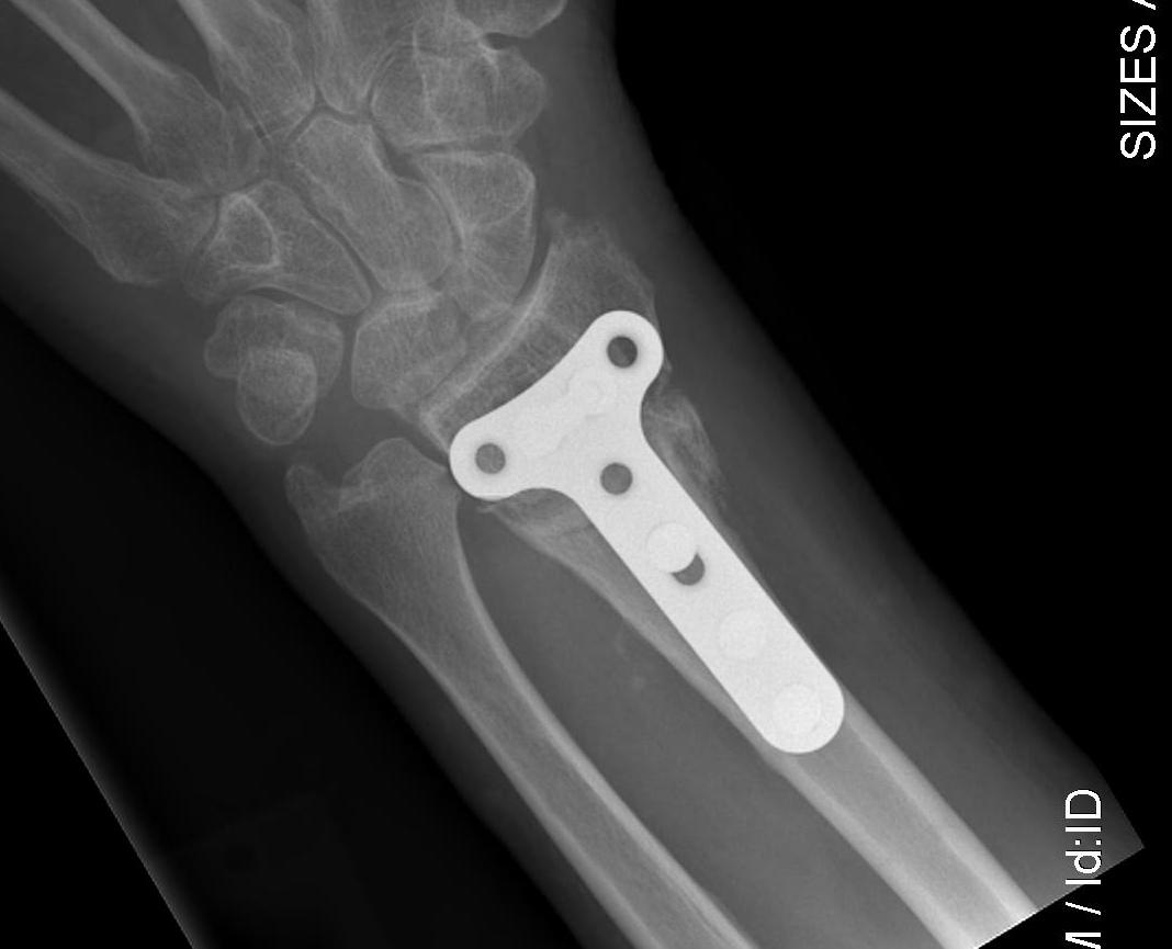
Volar opening wedge
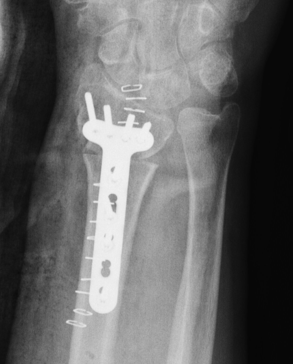
Advantage
Volar approach and plate
Disadvantage
May require dorsal approach to bone graft
Technique
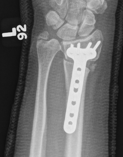
Vumedi volar osteotomy for distal radius fracture
Bed of FCR approach
- release brachioradialis
- protect structures with retractors
- perform osteotomy parallel to articular surface
- sufficient distal bone for screw fixation
- correct distal radius in two planes
- apply volar plate
- bone graft defect through radial aspect of wound
+/- dorsal approach to insert bone graft
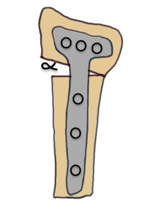
Dorsal opening wedge osteotomy

Advantage
Lengthens the distal radius
May be easier to correct in coronal and sagittal plane
Disadvantage
Dorsal approach / dorsal plate - extensor tendon issues
Technique
3 / 4 dorsal approach
- expose distal radius
- can use half pins to control distal fragment
- protect structures with homan retractors
- osteotomy with microsagittal saw
- correct radial articular surface in sagittal & coronal planes
- trapezoidal bi-cortical iliac crest autograft / synthetic graft
- dorsal locking plate
Distal radial osteotomy & ulnar shortening
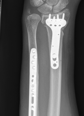
Indications
Unacceptable radial alignment
DRUJ not reduced by radius osteotomy
DRUJ articular surface acceptable
Distal radial osteotomy & ulnar resection / fusion
Indications
Unacceptable radial alignment
DRUJ not reduced by radius osteotomy
DRUJ articular surface unacceptable
Options
Bower's hemiresection
Darrach's
Suave-Kapandji
Intra-articular osteotomy
Indication
Step deformity
No radiocarpal osteoarthritis
Technique
Dorsal 3/4 approach and open radiocarpal joint