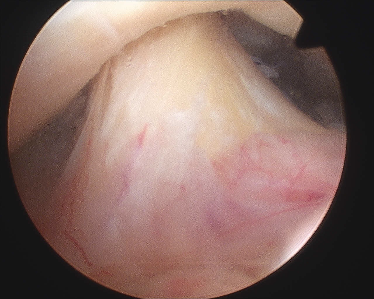
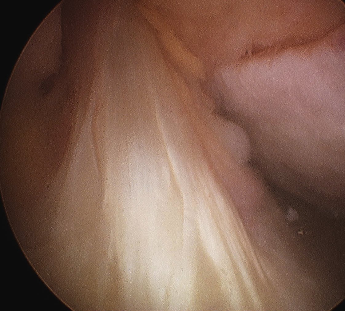
Arthroscopy

Arthroscopy

Position


1.  Lateral decubitus

- stabilise patient with beanbag or lateral rests

- apply skin traction to forearm

- place traction pole at foot of table opposite surgeon

- suspend arm with 10 lb weight

- abduction 60°

- forward flexion of 20°

- tilt top shoulder posteriorly 30° so that glenoid is parallel wwith bed

- mark bony landmark

- prep & free drape


Osteoarthritis

Non Operative Management


Options / ELM POPI


Education

Lifestyle Management

- weight loss

- reduce sport

Physiotherapy

Orthotics

- walking stick

- braces

Pharmaceuticals

- NSAIDS

- acetominophen

- glucosamine

Injections

- cortisone

- hyaluronic acid


Background

DefinitionKnee OCD MRI Cartilage Intact


Osteochondritis Dissecans

- separation of avascular fragment of bone & cartilage


Epidemiology


M : F = 2:1


Mean age 18 years

- can present as young as 9


4:1000


Background


ACL Normal ArthroscopyACL Normal Arthroscopy


Anatomy


Developmental Anatomy


Knee joint first appears as a mesenchymal cleft at 8 weeks gestation

- ACL and PCL separate entities by week 10