Definition
Abnormal posteriorly directed sagittal plane curve of spine
Scoliosis Research Society
Thoracic
Normal range thoracic kyphosis is 20-40°
- measured over T1 to T12 by Cobb method
- upper limit of normal thoracic kyphosis < 45°
Cervical & Lumbar
- lordosis is normal
- any kyphosis (>5°) considered abnormal
Classification Scoliosis Research Society
Postural
Scheuermann's Disease
Inflammatory / Ankylosing Spondylitis
Congenital
- failure of segmentation / formation / mixed
Iatrogenic
- post laminectomy / tumour excision in child / radiotherapy
Traumatic
- acute fracture / anterior wedging
- chonic - osteoporosis, OI
Infection
- TB
Metabolic
- Osteoporosis
- OI
- Mucopolysaccharidoses
Neuromuscular
- Polio
- Spinal muscular atrophy
- UMN Syrinx
- SB
Developmental
- Achondroplasia
- SED
- morquio's
Postural Kyphosis
Often confused with Scheuermann's
Examination
Gradual, no angular curve
Patient can voluntary correct roundness on stance
Prone hyperextension test
- reversal of thoracic spine hyperkyphosis
X-ray
No structural vertebral changes
Corrects on supine xray on bolster
Management
No treatment necessary
Post - Laminectomy Kyphosis
Mechanism
Occur because posterior supporting structures removed
- normally resist gravity producing kyphosis
Adult
Following radical laminectomy
- facet joints removed bilaterally
Infection post surgery
Growing child
Usually after excision spinal cord tumour
- radical laminectomy removing facet joints bilaterally
Management
Laminectomy
- prevention is key
- must preserve at least 1/2 of each facet joint or one whole facet / level
- if not possible, fusion indicated
Child
- must recognise potential for deformity & closely observe child
- orthoses don't often work
- if deformity develops & progresses, fusion usually indicated
Post-Traumatic Kyphosis
Risk Factors
Wedge fracture with initial kyphosis of > 30o
Focal kyphosis may develop if there is damage to the anterior column
- worse if posterior column fracture as well
- Most common TL junction
Indication for surgical intervention
Neurological deficit due to kyphosis
Refractory pain
Progress of deformity
Poor cosmesis
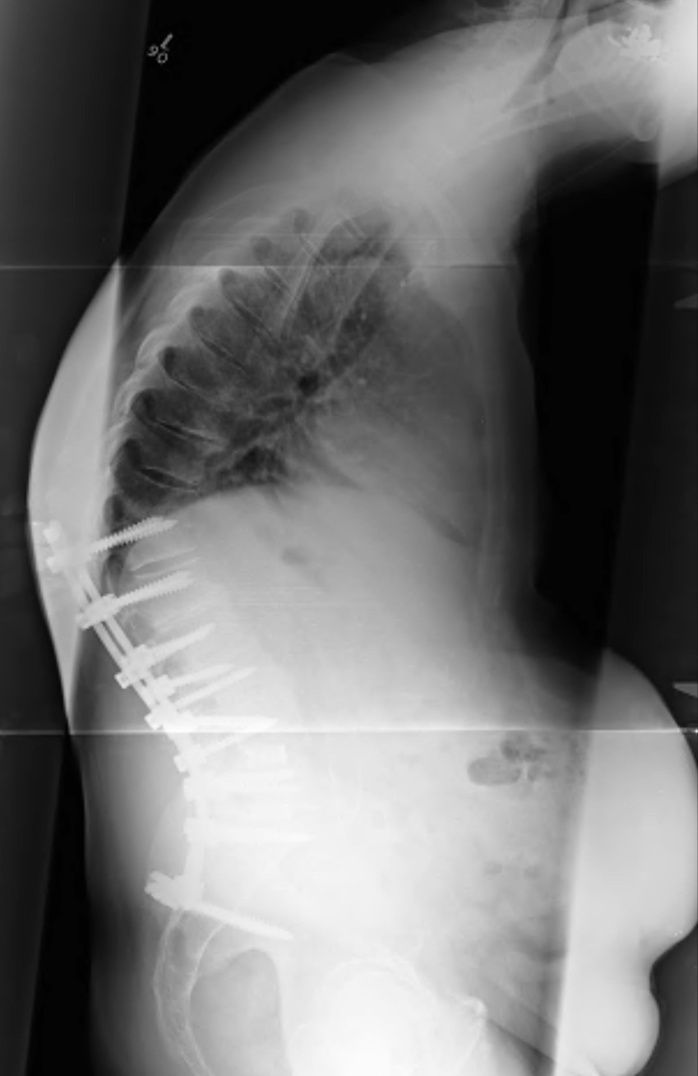
Management
If curve < 60°
- posterior instrumentation & fusion
If curve > 60°
- anterior approach usually necessary to obtain releases