HAGL
Definition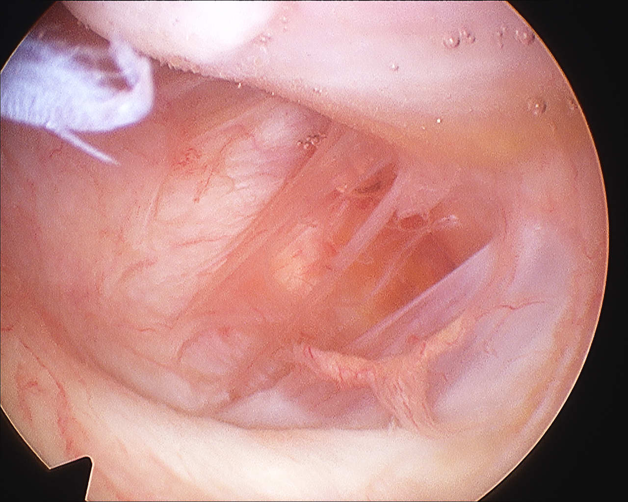
Humeral Avulsion of Glenohumeral Ligament
Incidence
Bokor et al JBJS Br 1999
- 514 cases surgical treatment traumatic instability
- incidence 7.5%
- 25% associated SSC tear
- likelihood of HAGL if no Bankart or MDI 27%
Humeral Avulsion of Glenohumeral Ligament
Bokor et al JBJS Br 1999
- 514 cases surgical treatment traumatic instability
- incidence 7.5%
- 25% associated SSC tear
- likelihood of HAGL if no Bankart or MDI 27%
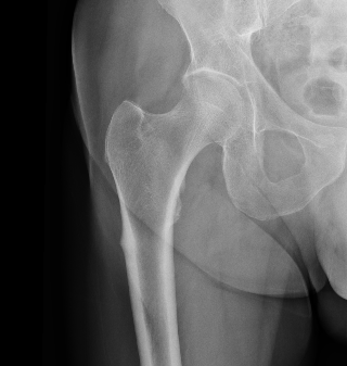
Subtrochanteric
Lateral femoral shaft
Stress fractures
Associated with long term bisphonate use
Black et al. NEJM 2020
- reduction of hip fracture risk v risk of atypical femur fracture
- 10 year period
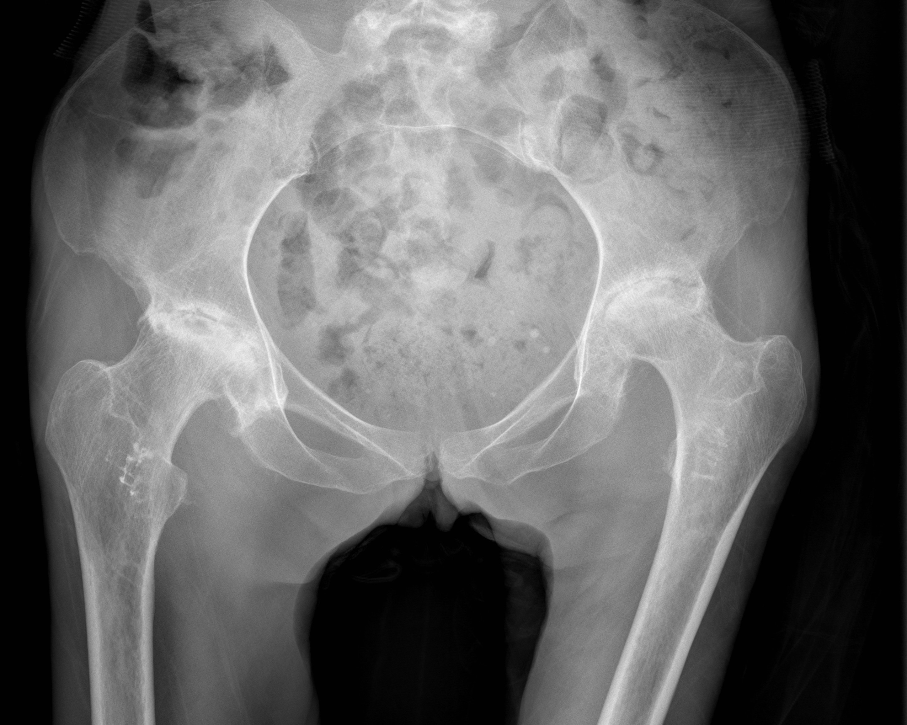
Self limiting syndrome of unknown aetiology
- hip pain associated with osteoporosis of proximal femur
AVN
- AVN of the hip in pregnancy is rare but possible
- TOH tends to be diffuse on MRI, while AVN is localised
- extends to neck and metaphysis
- transient osteoporosis has normal bone scan
Rare
- M: F 3:1
Non-traumatic or traumatic condition of femoral head with bone death
20 - 50 yo (average 38)
- M: F 4:1
70-80% with AVN will progress within 1 year
Largely related to degree of displacement
Hawkins Type I
- 0% to 13%
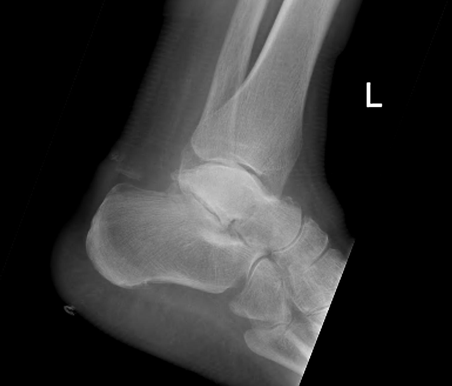
Hawkins Type II
- 20% to 50%
- usually only patchy and not a problem (rarely collapses)
Disorder of immune system
- antigen-antibody complexes
- stimulate release of proteolytic enzymes
- leading to vasculitis, synovitis and cartilage destruction
Articular Effects
- synovitis
- ligamentous and capsular laxity
- cartilage destruction
- osseous erosion
Vasculitis
- rheumatoid nodules
Complain of pain with prolonged standing
Complain feet tire easily
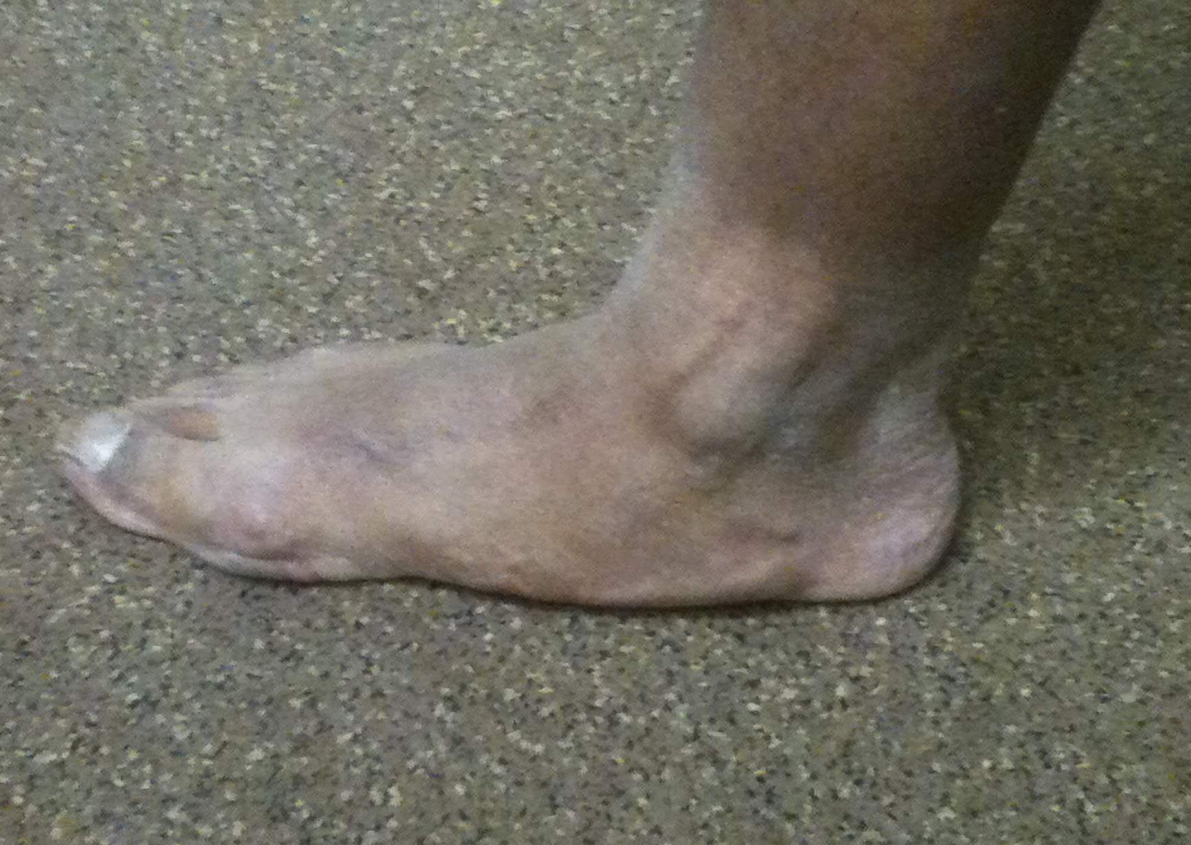
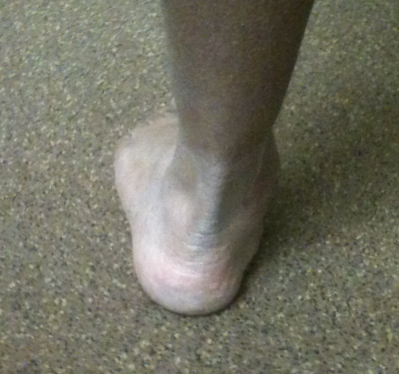
Overall alignment
Heel raises
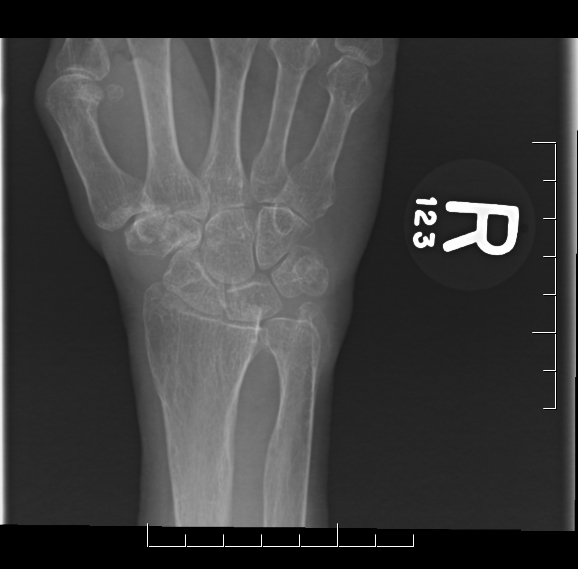
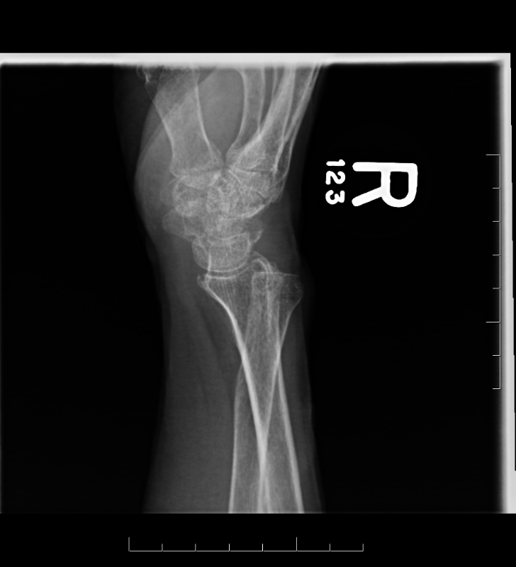
Young men
FOOSH
- axial load, dorsiflexion and radial deviation
DISI occurs in ulna deviation
Type A Stable acute fracture
A1 Tubercle
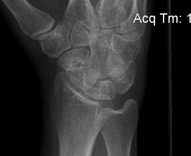
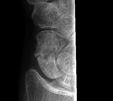
Convincing association with development of osteoarthritis
- arthritic changes beginning at radial styloid
- progress to scaphocapitate & capitolunate
Dorsal
- most common
Volar
1. Acute traumatic peripheral tear TFCC with DRUJ dislocation
- usually major trauma
- dorsal or volar
2A. Distal radial fracture