Types
Dystrophic or Metastatic
1. Metastatic
- hypercalcaemia / hyperphosphatemia
2. Dystrophic
- more common
- onto damaged connective tissue
- tendon / ligament / cartilage
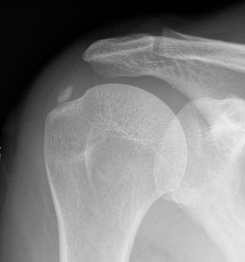
- calcific tendonitis shoulder
- Pellegrini-Steida lesion MCL
Pathology
Deposited around chondrocytes & into avascular portion of CT
- crystals grow by accretion
- early on like cream
- later like chalk
- can be inert or surrounded by inflammatory reaction
- crystal shedding into joint causes synovitis
Clinical Features
Two syndromes
1. Acute or Subacute Periarthritis
Pain near a large joint
- not intra articular
- after minor trauma
- warm & swollen tendon / ligament
- calcific tendinitis rotator cuff
- Pellegrini-Stieda lesion MCL
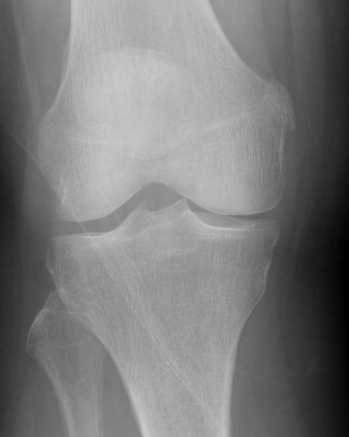
2. Chronic Destructive Arthritis
HA crystals found in association chronic erosive arthritis
- unknown if cause or effect
- destructive arthropathy seen in shoulder with cuff arthropathy
- i.e. Milwaukee shoulder
- whether related to HA unknown
Aspiration
Crystals too small to be seen with light microscopy
- hence will not see on aspiration
X-ray
Periarthritis seen as calcium in tendon
- especially rotator cuff
- chronic HA arthritis doesn't show on xray as well as CPPD
Management
Non operative
Periarthritis
- RICE
- NSAID
- HCLA
Operative
Surgical removal
- calcific tendonitis
- problematic Pellegrini Steida
Chronic Arthritis
- treat as OA
- early arthroplasty if rapid bone destruction