Definition
Injury to ulnar collateral ligament of thumb MCPJ
Interferes with pinch grip and grasp and thumb is ineffective as a post

Aetiology
Valgus / forced abduction
10 x more common than injuries to radial collateral ligament
Gamekeeper's thumb's
- secondary to repetitive breaking pheasant's neck
- chronic injury
Skier's thumb
- acute injury
Anatomy
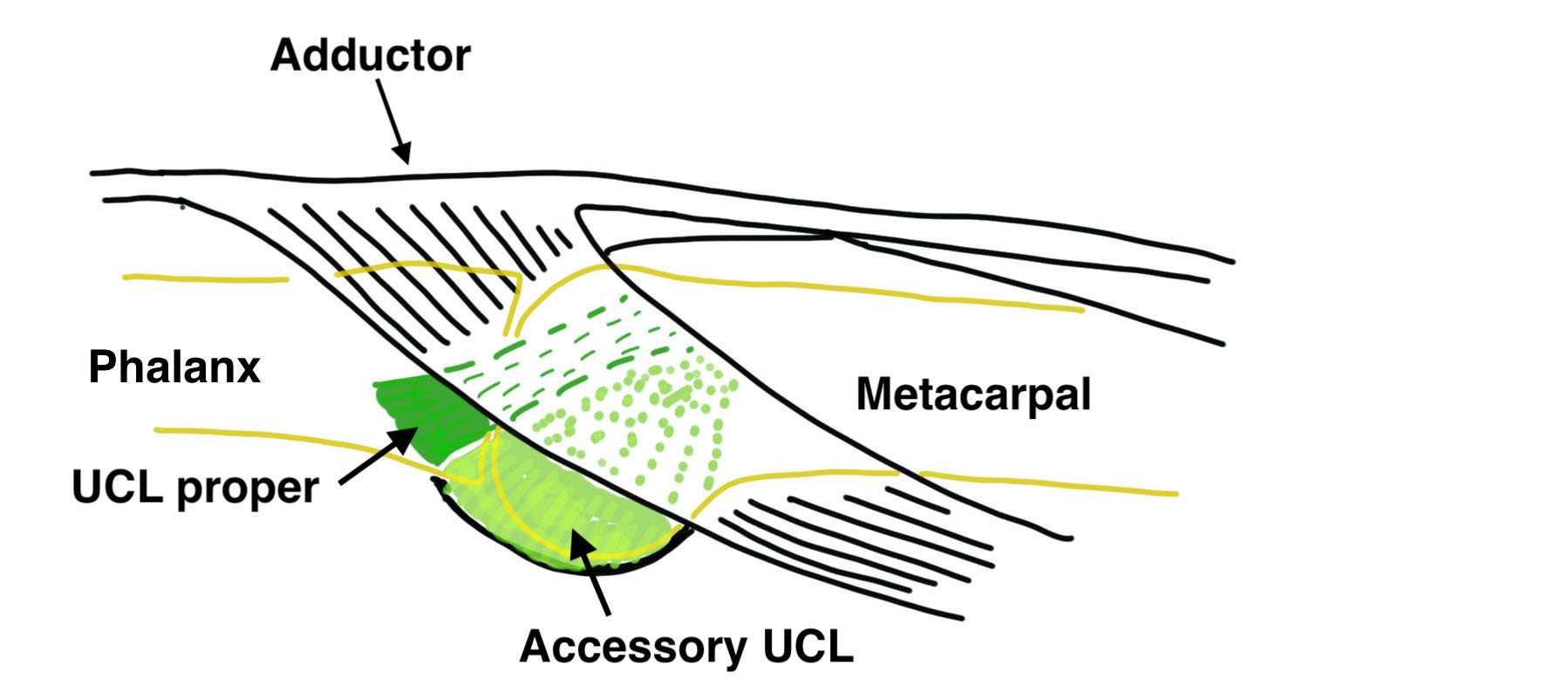
Ulna collateral ligament
Proper
- origin dorsal metacarpal head
- passes to volar aspect proximal phalanx
Accessory
- volar side of proper ligament
- attaches to volar plate
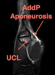
Adductor aponeurosis
Superficial to ulna collateral ligament
- inserts into ulna border thumb extensor mechanism
- via the ulna sesamoid
Pathology
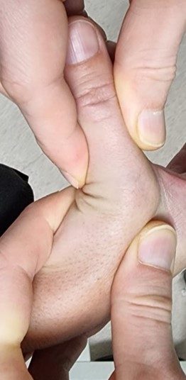
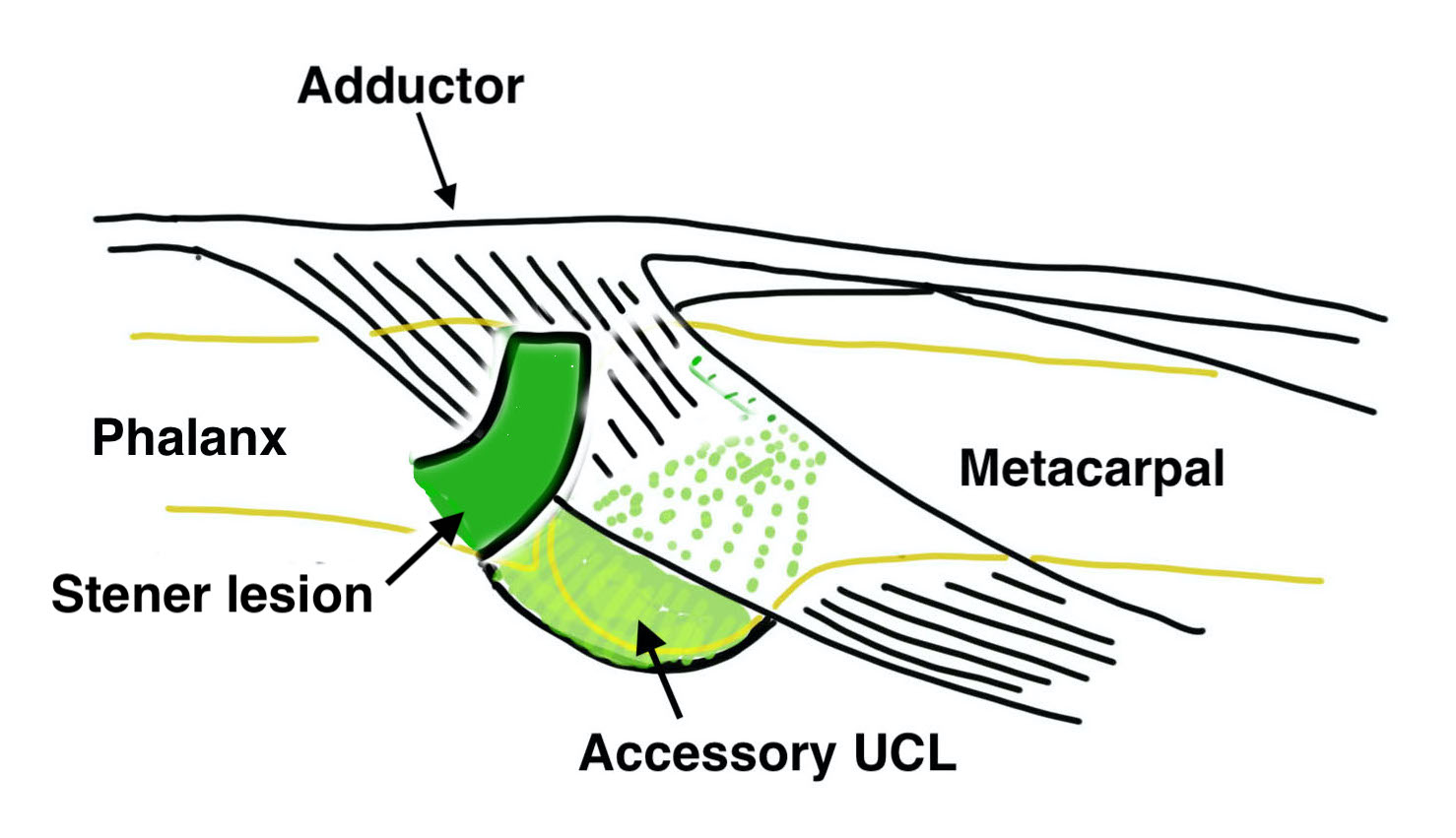
Stener lesion
- distal end of UCL flipped superficially over adductor aponeurosis
- will not heal
- may be able to palpate a lump
- use MRI to diagnose
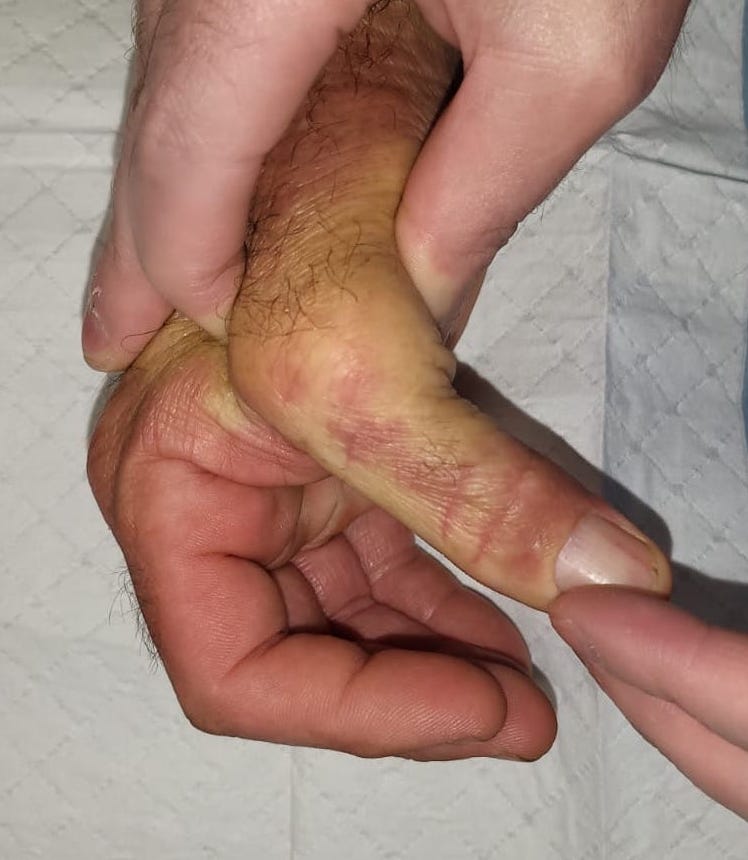
Examination

Painful, swollen MCPJ
Tenderness along UCL
Abduction Stress Test
In full extension and 30° compared to other side
- increased opening at 30o - injury to ulna collateral ligament proper only
- increased opening at 30o and full extension - injury to both accessory and ulna collateral ligament
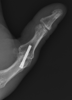
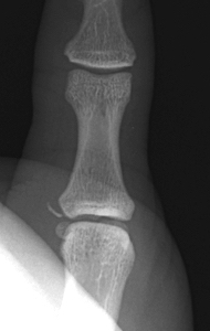
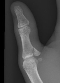
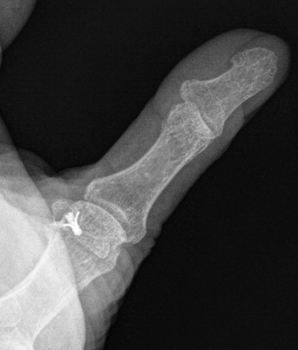
X-ray
Bony avulsion
1. Small fragment pulled away from proximal phalanx
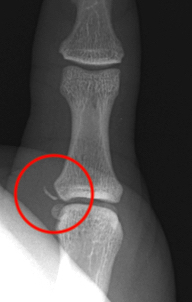
2. Large intra-articular fracture involving >1/4 articular surface

3. Salter Harris III in pediatric population
MRI
Anatomy
A. Undisplaced
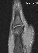
Distal tear of ulna collateral ligament on coronal MRI
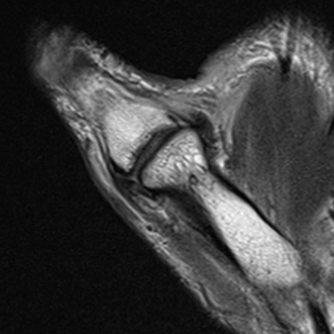
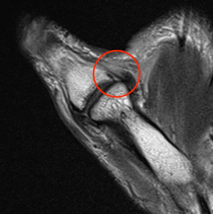
Distal tear of ulna collateral ligament on coronal MRI
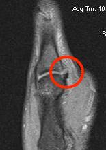
B. Displaced UCL
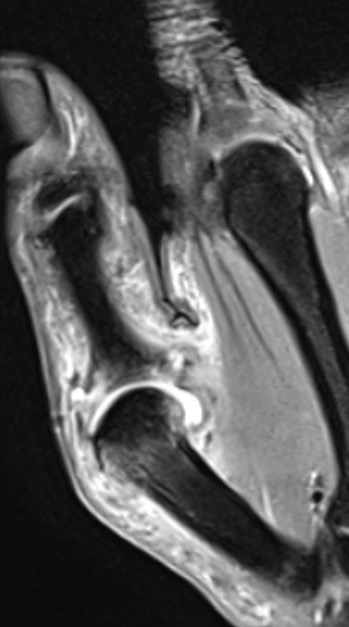
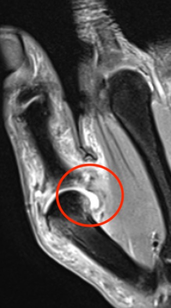
Coronal MRI demonstrating displaced distal UCL avulsion
Management
Non operative
Indications
Partial tear
Undisplaced complete tear
Undisplaced bony fragment
Management
6/52 thumb spica
Operative
Indications for surgery
Stener lesion
Complete tear with displacement
Displaced bony fragment
Salter Harris III
Chronic injury with instability
Acute injury
Results
Milner et al J Hand Surg Am 2015
- 43 cases acute UCL injury with MRI
- all partial tears / minimally displaced / displaced < 3 mm healed with immobilisation
- tears displaced > 3 mm failed immobilisation
- biomechanical study
- primary repair + suture tape augmentation > primary repair > ligament reconstruction
Gibbs et al Orthop J Sports Med 2020
- 18 thumbs in athletes
- primary repair of acute UCL injury augmented with suture tape
- average return to sport 5 weeks
Surgical technique
AO foundation surgical approach
Vumedi primary repair with suture anchor
Arthrex internal brace surgical technique PDF
Vumedi primary repair augmented with internal brace
Dorso-ulnar approach to the MCPJ of the thumb
- dorsal incision along ulna border MCPJ
- divide Adductor pollicis aponeurosis
- leave cuff for lateral repair
- identify UCL
Fixation options
- bony anchors for proximal or distal tears +/- internal brace
- direct repair of midsubstance +/- internal brace
- screw fixation of bony avulsions
Post operative management
- 6/52 thumb spica
Chronic Injuries
Options
Primary UCL repair +/- suture tape augmentation
Adductor advancement
Tendon reconstruction
MCPJ fusion
Results
Agout et al Ortho Traumatol Surg Res 2017
- 55 chronic injuries
- compared repair when able with reconstruction and fusion
- primary repair > fusion > reconstruction
Tendon reconstruction
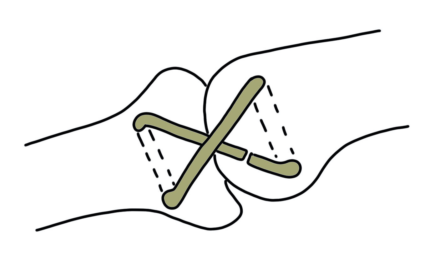
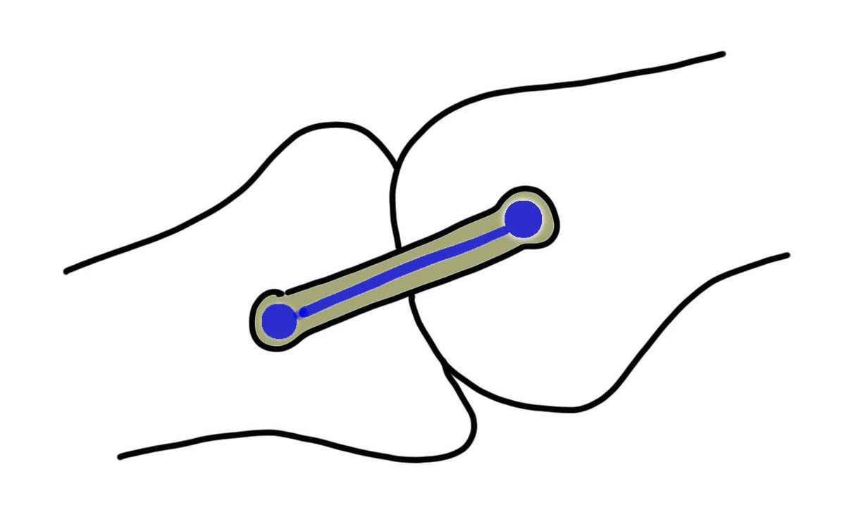
Figure 8 reconstruction Single bundle reconstruction with suture tape augmentation
Graft options
- palmaris longus
- strip of FCR if palmaris absent
- fourth toe extensor tendon
Technique
- figure of 8 through drill holes
- suture anchor fixation +/- suture tape augmentation
Vumedi tendon reconstruction with suture anchors and suture tape augmentation
MCPJ fusion