Normal Anatomy
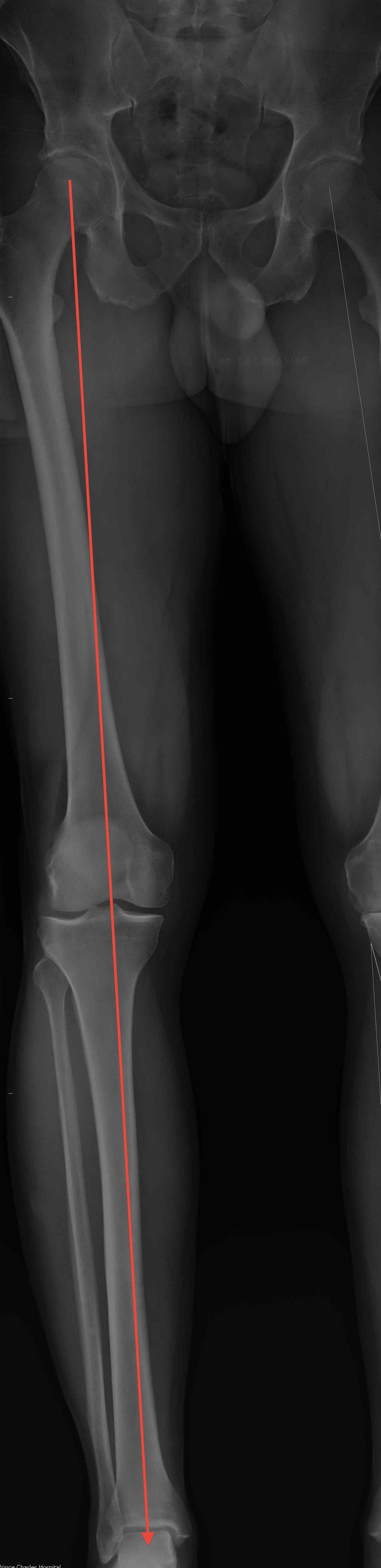
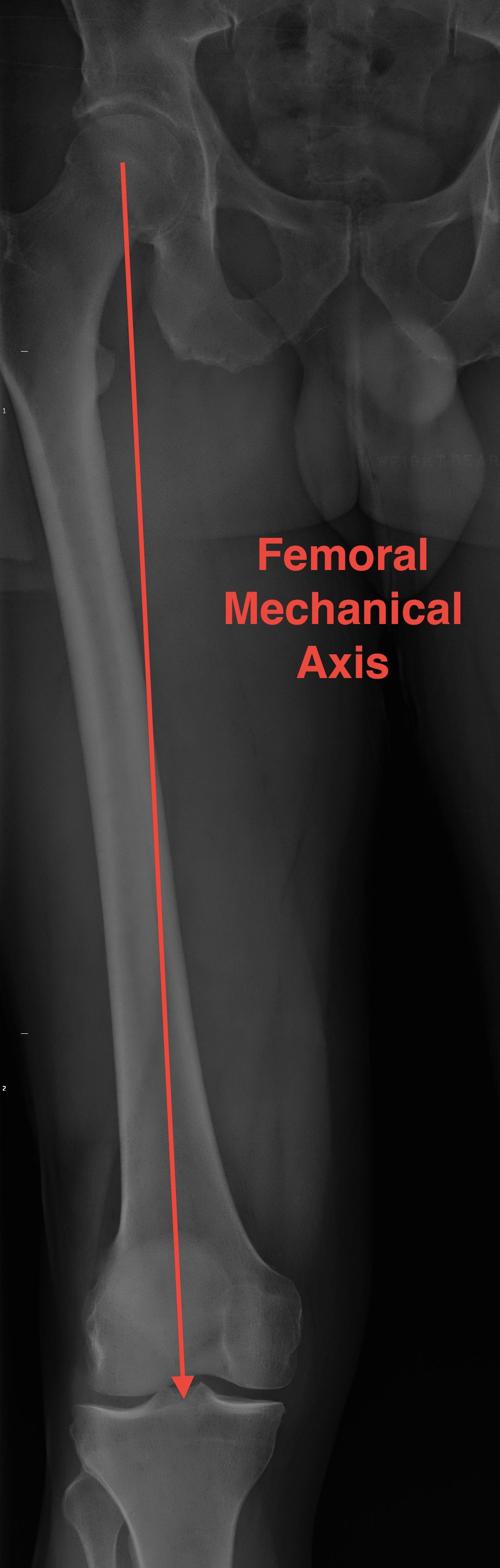
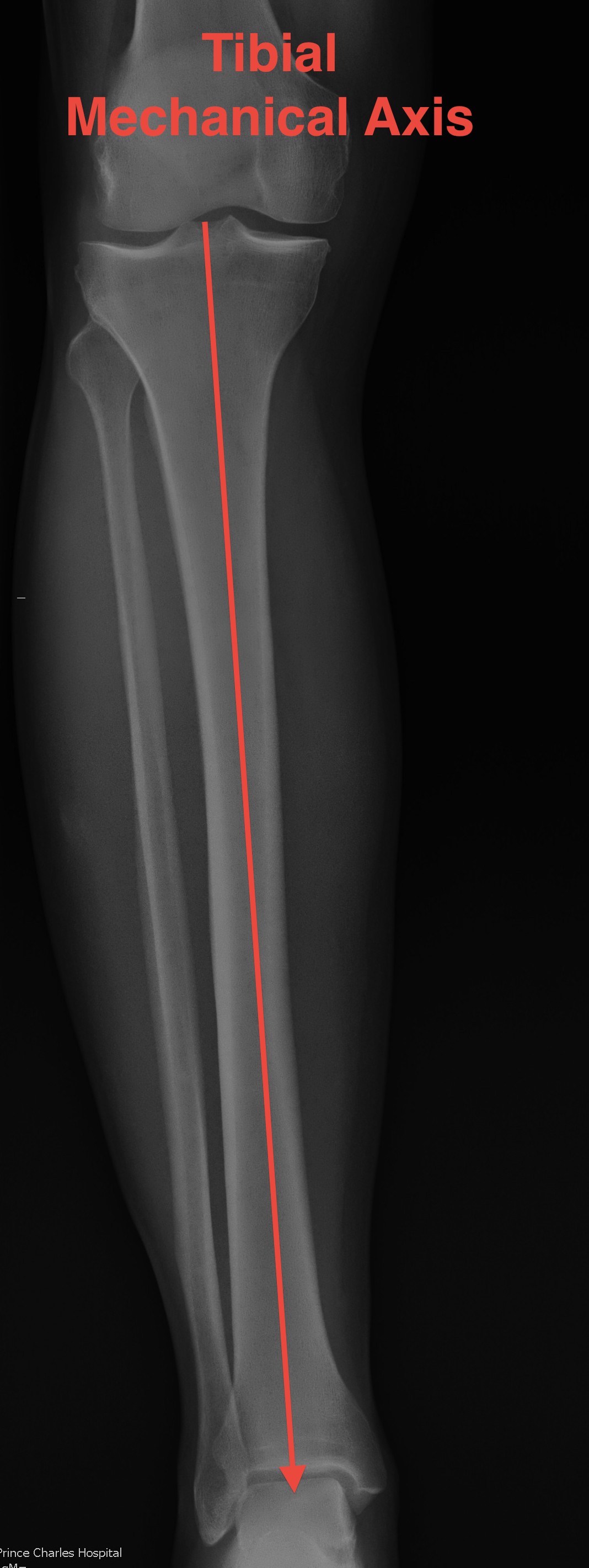
Mechanical Axis
| Coronal mechanical axis | Femoral mechanical axis | Tibial mechanical axis | Sagittal mechanical axis |
|---|---|---|---|
| Center femoral head to center of ankle | Center of femoral head to center knee | Center plateau to center of talus / ankle | Center of femoral head to center of ankle |
|
10mm medial to the frontal plane center Medial tibial spine |
Just anterior to center of knee joint Aids in passive locking of knee joint in full extension |

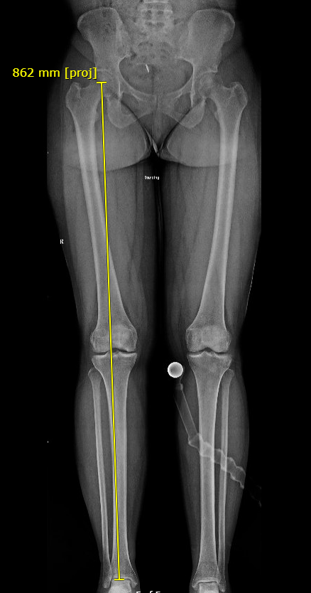
Varus malalignment left knee Valgus mechanical axis right knee
Mechanical Axis Deviation
- distance from center of knee to MA in mm
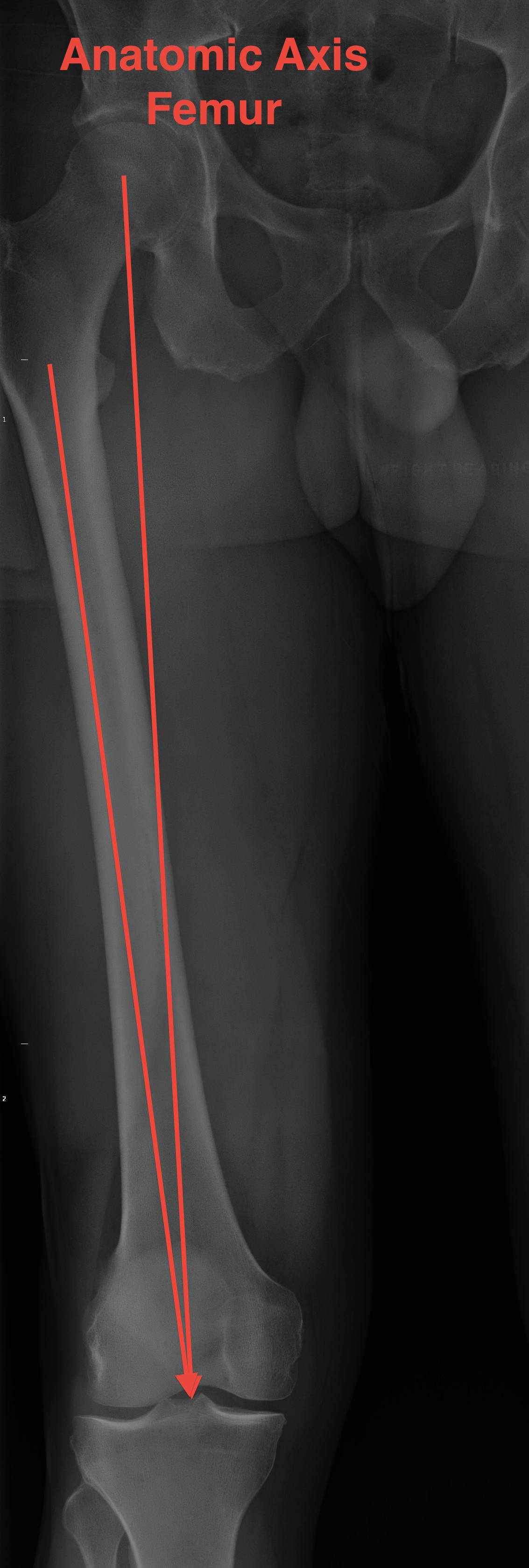
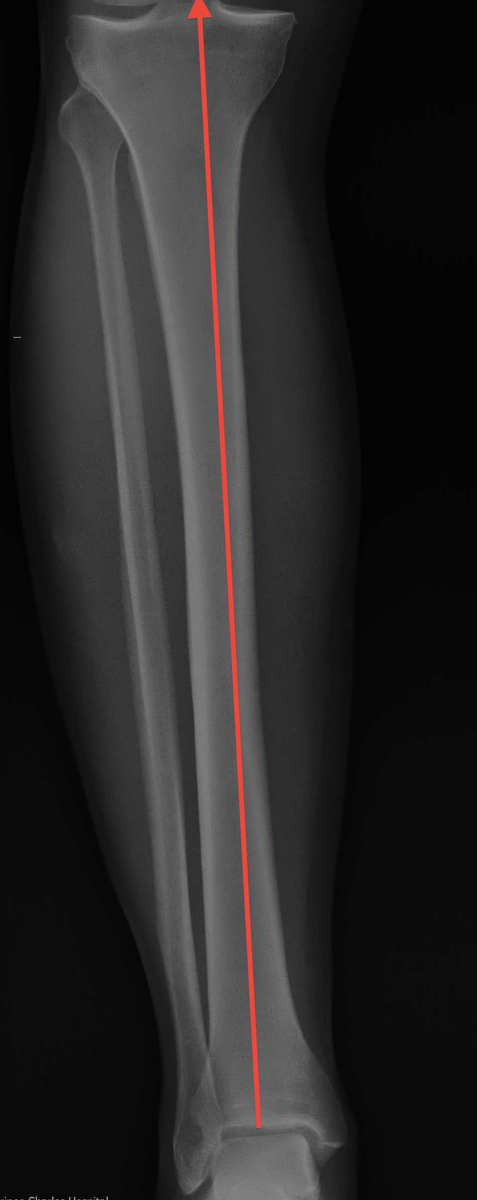
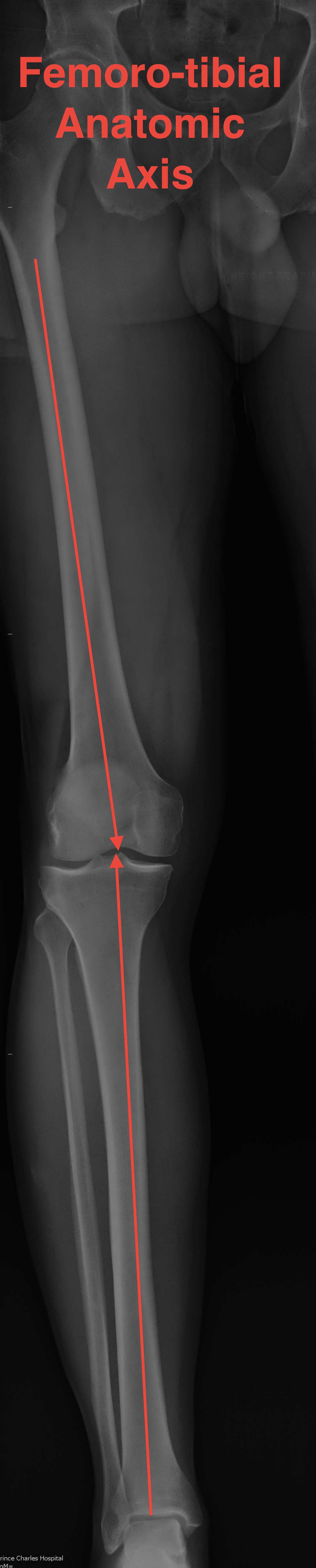
Anatomical Axis
| Coronal anatomical femoro-tibial axis | Femoral anatomical axis | Tibial anatomical axis |
|---|---|---|
|
Center of femoral shaft to center of knee to center of tibia |
Piriformis fossa to center knee joint | Center plateau to center of talus / ankle |
|
5-7o valgus - increased in shorter femurs - decreased in longer femurs |
6o from mechanical axis |
Knee
| Knee joint |
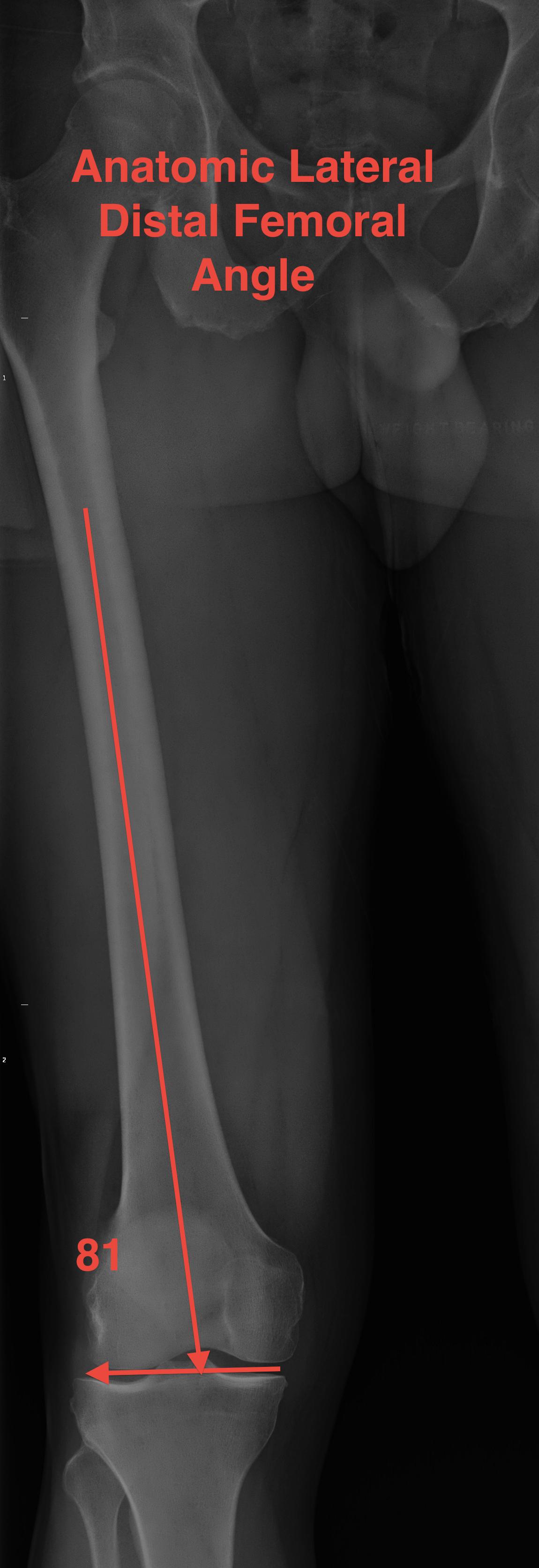
aLDFA (anatomical Lateral Distal Femoral Angle) |
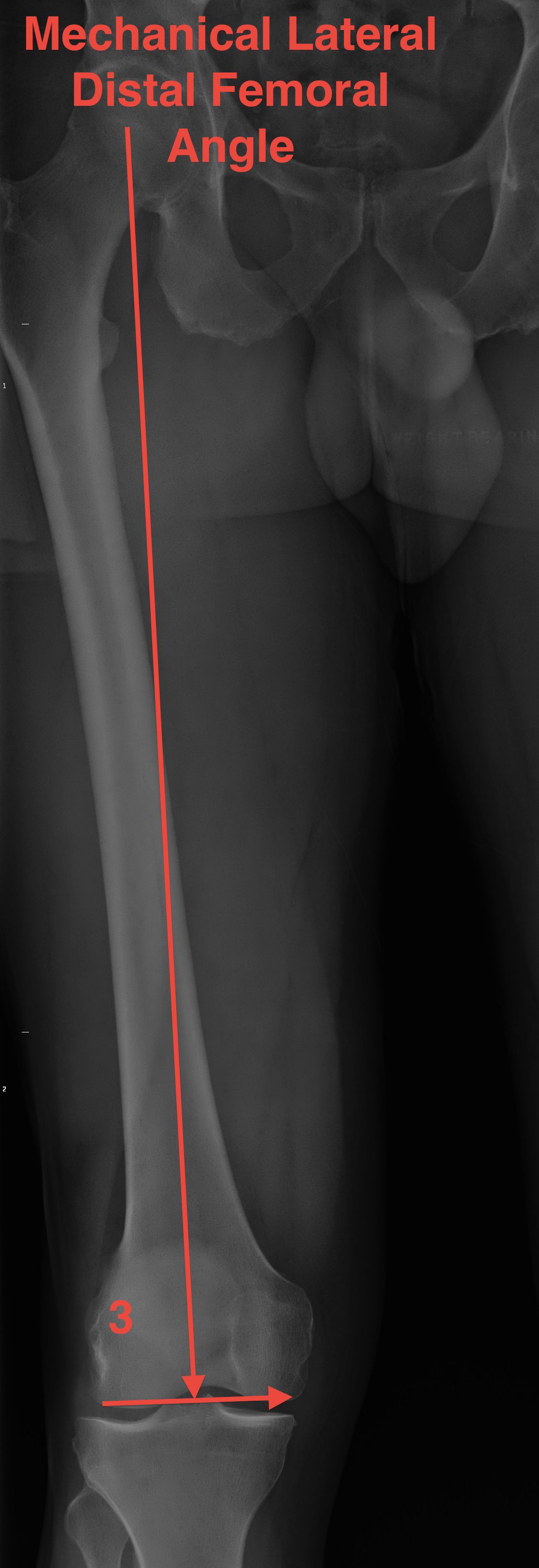
mLDFA (mechanical Lateral Distal Femoral Angle) |
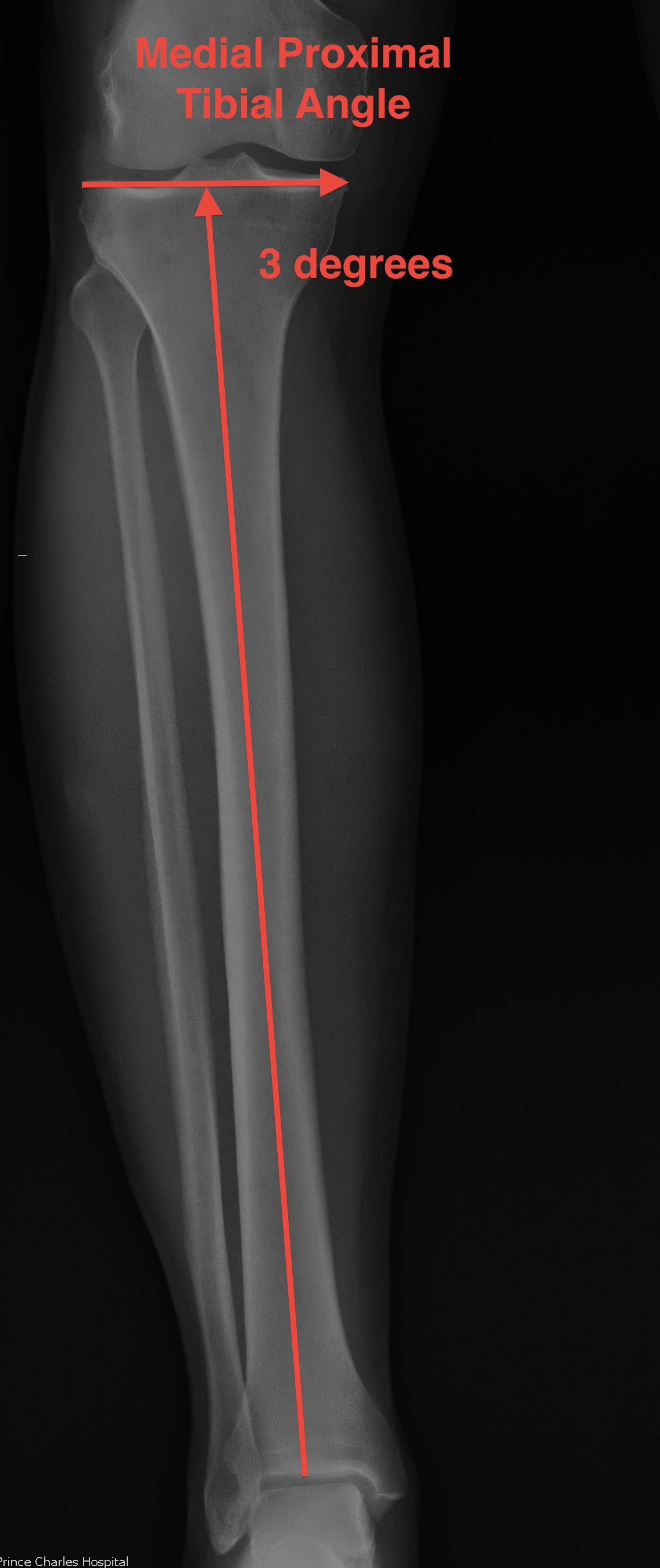
MPTA (medial proximal tibial angle) |
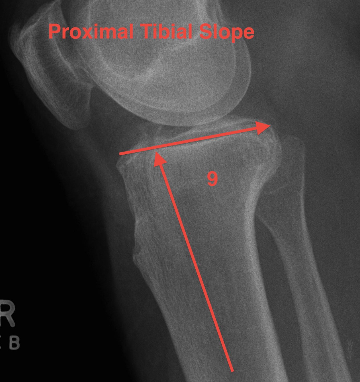
Posterior slope |
|---|---|---|---|---|
| 3o valgus relative to MA | 81o | 87o | 87o or 3o varus | 9° |
| 6o valgus distal femur | ||||
| 3o varus proximal tibia |