 Incidence
10 -15%
Include
- marginal necrosis
- wound slough
- sinus tract formation
- dehiscence
- haematoma
- oozing knee wound
Blood supply
Anterior knee has no muscles to supply vessels directly
- dermal plexus
- any subcutaneous dissection disrupts this & potentiates necrosis
- any skin flaps raised must be below subcutaneous fascia
Blood supply comes across medially
Prevention
1. Avoid closely parallel scars
- 7 cm bridge is minimum
- use most lateral of 2 incisions
2. Gentle tissue handling
3. Avoidance of undermining & thin skin layers
4. Careful closure of deep layer
- watertight, prevents oozing
5. Closure with knee in flexion
6. Avoid CPM
- >40° on first 3 days post-op associated with decreased oxygen tension
7. Lateral Release
- decreased skin O2 tension
- attempt to preserve SLGA
Concerns preoperatively
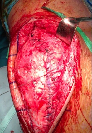
1. Consult plastic surgeon
2. Sham incision
3. Tissue expanders
4. Pre-op flap
- pedicled medial gastrocneumius flap + SSG
Patient Related Factors
1. Steroids
- decreased fibroblast proliferation necessary for wound healing
2. RA
3. DM
4. Obesity
- exposure, fat has poor and tenuous blood supply
5. Malnutrition
- Alb<3.5gm/L
- lymphcytes <1500 cells/L
6. Smoking
7. Chemo
- MTX slight inc in wound healing problems? others none
8. Hypovolaemia
Continuous Haemo-Serous drainage
Prolonged drainage
- 17-50% eventually become culture proven infection
Early management
- immobilise in splint
- local wound care
- cease anticoagulation / LMWH / Aspirin / NSAIDS
- trial vac dressing (drains haematoma / keeps sterile)
Drainage
- timing debatable
- rationale is that the wound in this situation is not a closed system
- that bacteria are entering the wound while it is still draining
- maybe better to reopen wound and debride it
- commonly find subcutaneous or deep haematoma as cause
Non-Draining Haematoma
- no evidence to support drainage
- drain if causing excessive soft tissue tension or restriction of motion
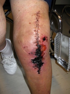
Superficial Soft Tissue Necrosis
Management
- aggressive surgical debridement & closure
- < 3cm in diameter should heal
- > 3cm needs formal debridement and skin closure with SSG, flap etc
Full Thickness Soft Tissue Necrosis
Metal on view
- necessitates immediate, extensive debridement
- medial gastrocnemius flap