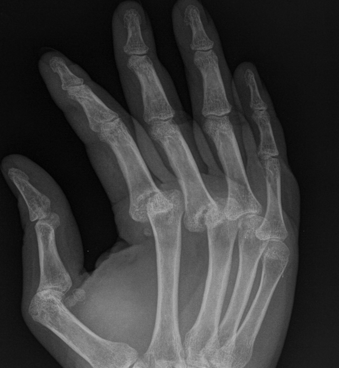
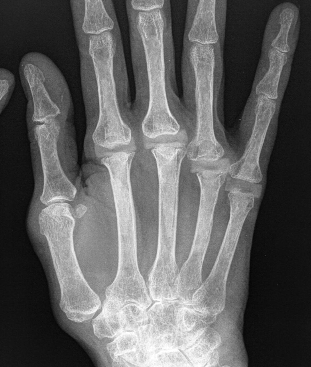
Deformity
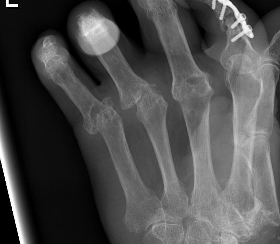
Ulna drift & volar dislocation
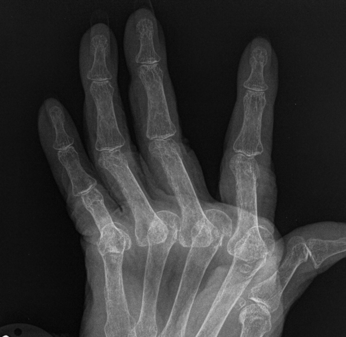
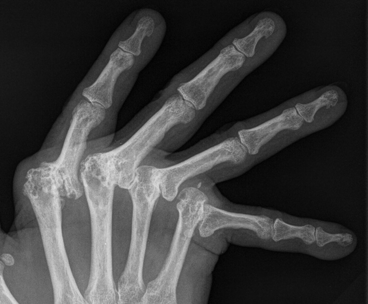
Causes of MCPJ Deformity
Ulna Drift / Ulna Dislocation
1. Physiological
- gravity
- lateral pinch pressure
- power grip
2. Anatomic
- shape of MC heads
- collateral ligament length & orientation
- intrinsics to LF asymmetric (hypothenars strong)
3. Pathological
- joint / capsule instability due to bony erosions
- collateral ligament stretching due to synovitis
- ulna/volar dislocation flexor tendons due to stretching pulleys
- ulna dislocation extensor tendons due to stretching sagittal bands
- intrinsic contracture
- radial deviation of wrist (Landsmere) redirecting line of pull of tendons
- volar / ulna carpal subluxation
Nalebuff Classification MCPJ
Stage I - Synovitis
- medical treatment and splinting
- synovectomy
Stage II - Synovitis + Ulna deviation
- medical treatment and splinting
- synovectomy + soft tissue reconstruction
Stage III - Moderate joint destruction / Volar subluxation
- soft tissue reconstruction possible
- arthroplasty gives more reliable results
Stage IV - Advanced joint destruction
- fixed joint deformities
- arthroplasty with soft tissue releases
Management
Stage I Synovectomy MCPJ
Indication
- marked synovial proliferation not responding to medical treatment
- 6/12 non-operative
- painful
- concern regarding progression to deformity
Contraindication
- joint destruction with articular erosion
- instability
- fixed deformity or dislocation
Technique
- incise hood on Ulna side extensor tendon
- make sure clear under volar plate & collaterals
Stage II Synovitis / Ulna Deviation / Preserved MCPJ
Synovectomy + Soft Tissue Reconstruction
1. Ulna side release
- divide transverse, oblique & sagittal bands
2. Crossed Intrinsic Transfer
- corrects ulna drift
- ulna side intrinsics are released
- transferred to the Ulna neighbour radial intrinsics
- reinsert through radial lateral band
- use EI for Index attach to radial side
- release EDM at little
3. Extensor Tendon Relocation
- ulna sagittal band release
- radial sagittal band tightening
Stage III / IV Destroyed MCPJ
Arthroplasty + ST Reconstruction as above
Swanson's Indications
- fixed or stiff MCPJs
- x-ray shows destruction or subluxation
- ulnar drift not reconstructable
- contracted intrinsic and extrinsics
- associated stiff IPJs
Swanson's contraindications
- infection
- inadequate skin coverage
- poor NV status
- irreparable intrinsic/extrinsic system
- insufficient bone stock
Aim
- painless joint with useful arc of motion
Results
ROM
- usually > 40°
- get about 10° improvement
Pain
- > 80% pain relief
- no increase in strength
Deformity correction
- up to 40% loss over time
- loss of correction often due to inadequate soft tissue balancing
Survival
- 90% 10 year survival
- silicon synovitis uncommon unlike for wrist or trapezial implants
Technique MCPJ Swanson Arthroplasty
Incision
- transverse incision dorsum
- full thickness flaps preserving dorsal veins
Dissection
- incise extensor hood on ulna aspect each joint
- may need formal intrinsic release but bony cuts may be enough
- incise and remove capsule and synovitis
MC head
- excise MC head with osteotome or nibbler sufficiently to accept implant
- with final cut at 90° to shaft
- this often means removing collaterals
- ream MC with awl or drill
PI
- do not resect P1 base
- just ream with awl
Trial
- resection of bone should allow no buckling of implant
- no impingement of MC on P1
- insert prosthesis proximal then distally
- should have passive motion of 90°
Soft tissue balancing
- ulnar intrinsic release
- crossed intrinsic transfer
- extensor tendon relocation