Aetiology
Knee forms from 3 separate compartments
Plica represents normal embryonic synovial septum that persists into adult life
Epidemiology
20% of knees have medial patellar plica at arthroscopy
Symptomatic plicae much less common 1-2%
Mean age 14
Types
1. Infrapatellar (ligamentum mucosum)
Most common / always asymptomatic
Role
- likely stabilises the fat pad to the knee
- may prevent fat pad impingement
2. Suprapatellar
Variable
- often incomplete
- may separate suprapatellar bursa from knee
- may hide loose body
2. Medial patellar
Least common / rarely symptomatic
Anatomy
- originate from medial wall of knee joint
- run obliquely to insert in medial infrapatellar fat pad
Occasionally symptomatic
- gets caught between patella & femur
Patient symptoms
- snapping / clicking
- medial pain
- may be able to palpate / reproduce symptoms
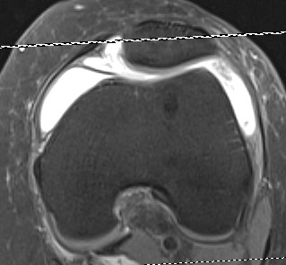
MRI
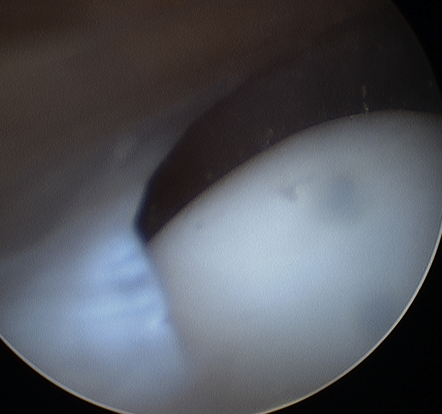
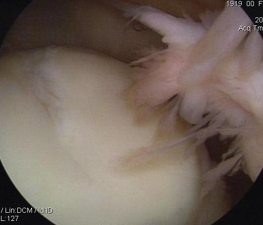
Arthroscopy
Normal
- thin
- no evidence inflammation
- no evidence chondral damage
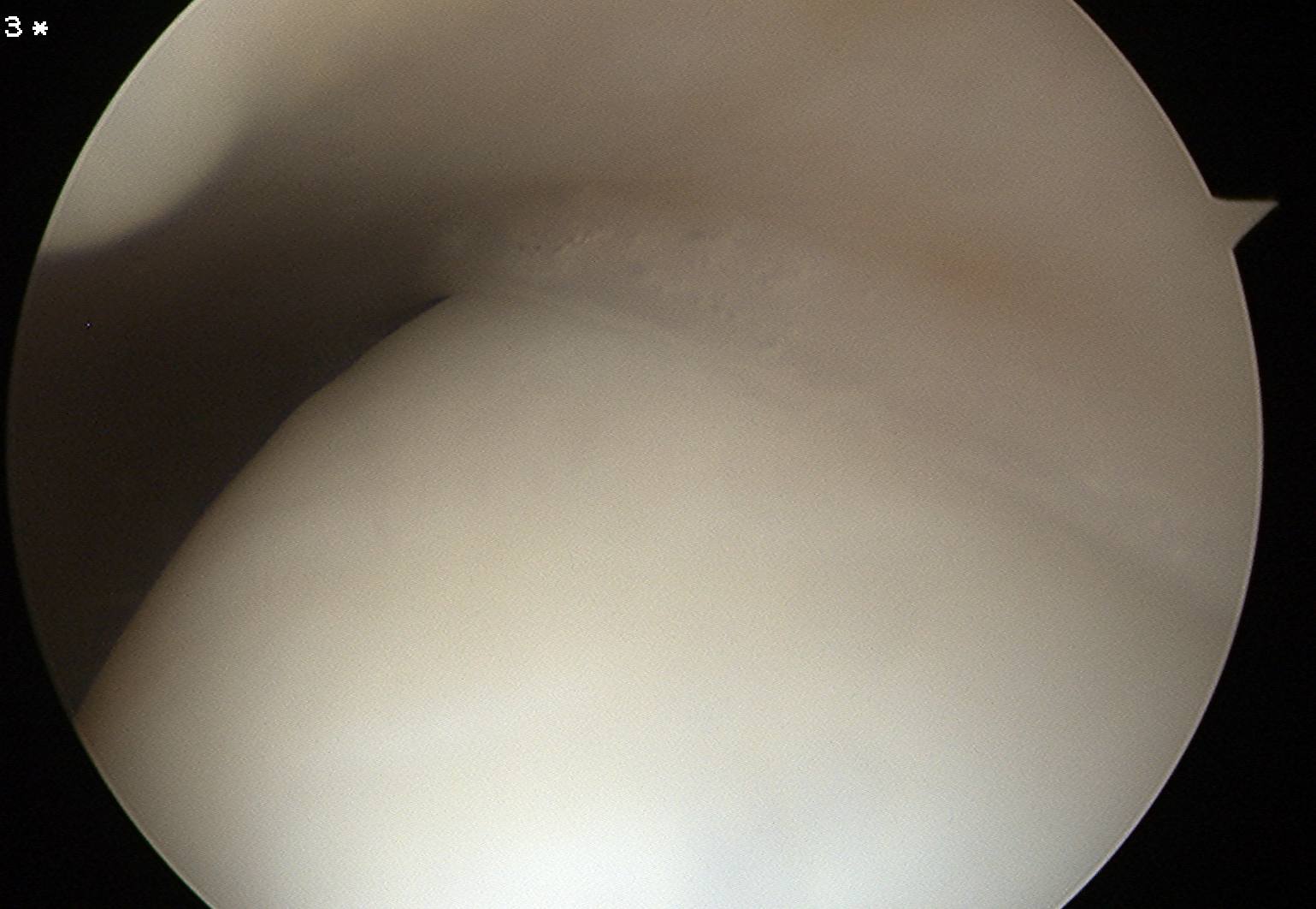
Abnormal
- thickened and inflammed
- obvious signs of chondral damage on MFC
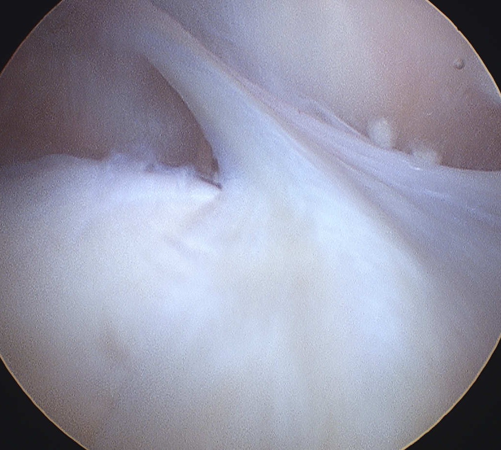
Arthroscopic resection
Divide plica to synovial membrane rather than completely excise