Look
Shoes
Walking aids
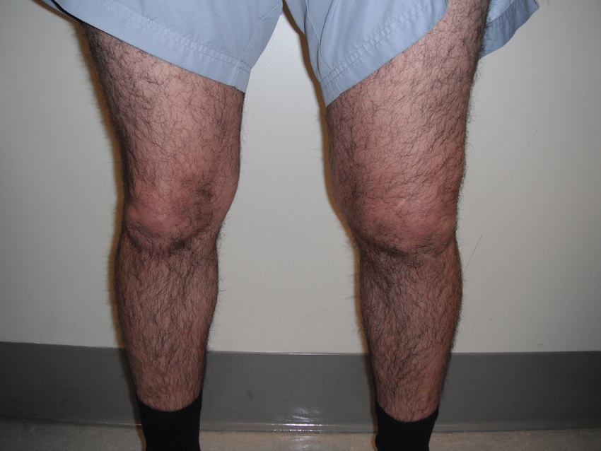
Front
Knee alignment
- physiological valgus
Patellar rotation
- squinting (inwards, increased PFA)
- grasshopper eyes (high and lateral)
Swelling
Quads Wasting
Scars
Side
Knee attitude
- flexion
- recurvatum
- push knees back
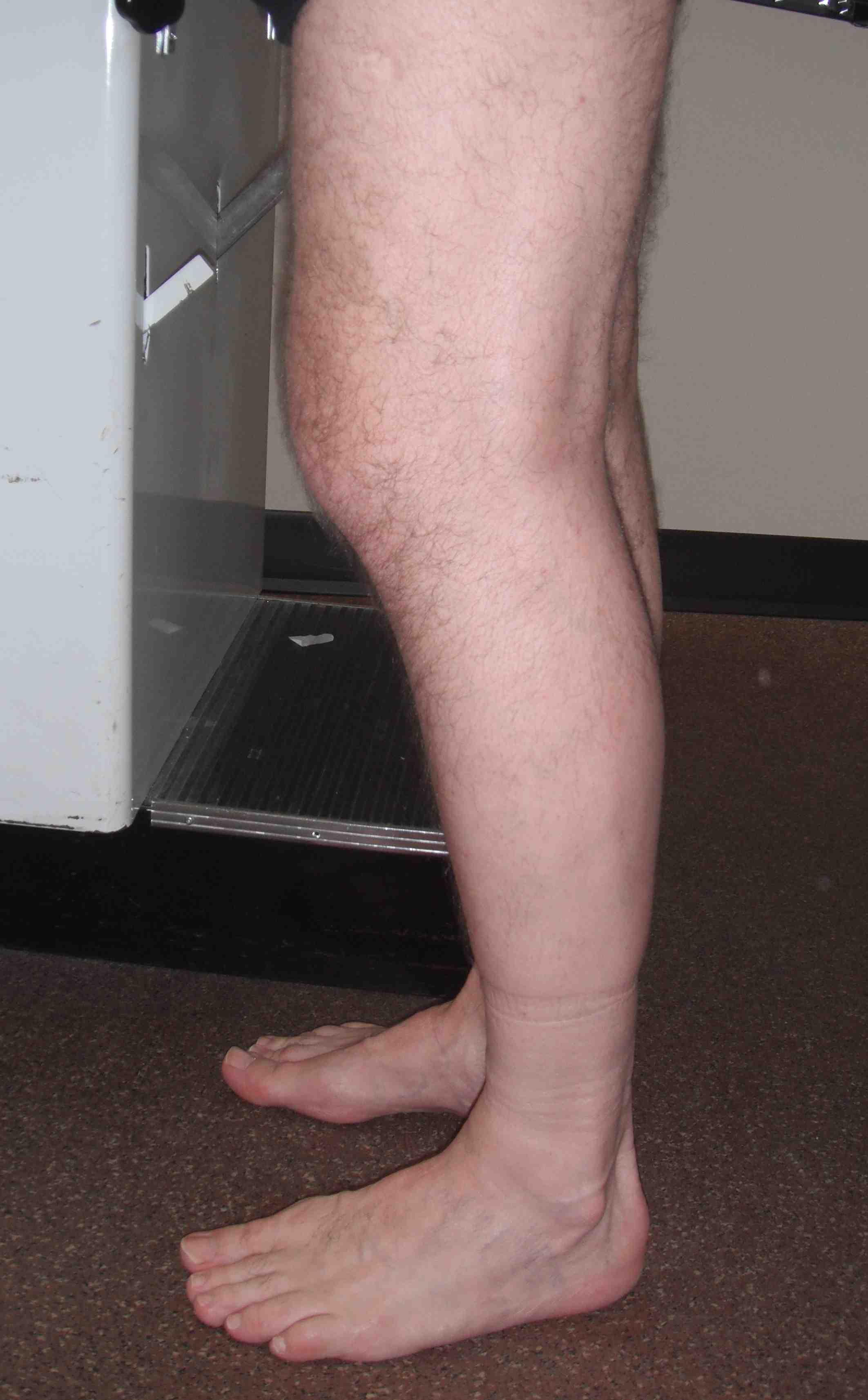
Step foot forward and bear weight
- examine arch
Scars
Behind
Hindfoot valgus
Swelling popliteal fossa
Wasting of hamstrings or calf
Level popliteal creases
Other Side
Knee attitude
- flexion
- recurvatum
- push knees back
Step foot forward
Scars
Gait
Rigid / Stiff
- decreased flexion / extension range
Antalgic
Weak knee
- back knee gait
Medial or lateral thrust
- valgus or varus moment about the knee
Foot progression angle
Sit on Edge of Bed
Patella tracking
- crepitus
J tracking
- patellar sharply deviates laterally in terminal extension
- or travel laterally until jumps into trochlea at midrange of flexion
Supine
Look
- quads wasting
- alignment
- scars
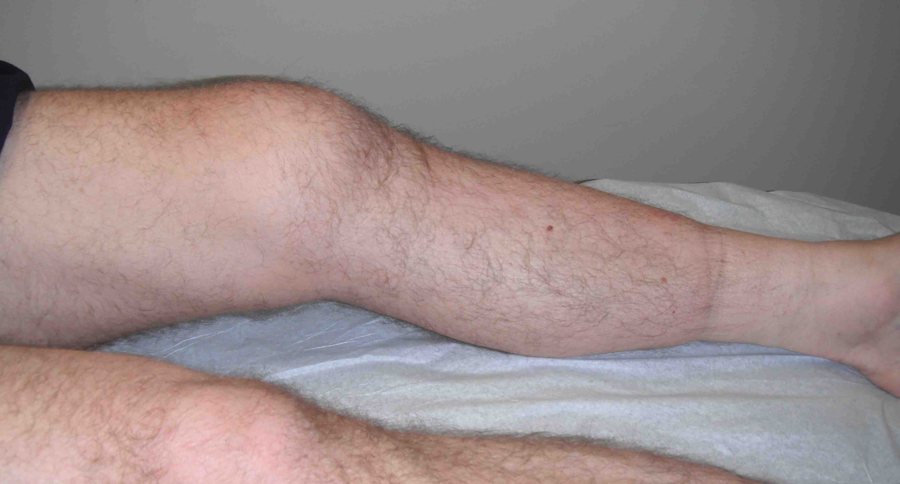
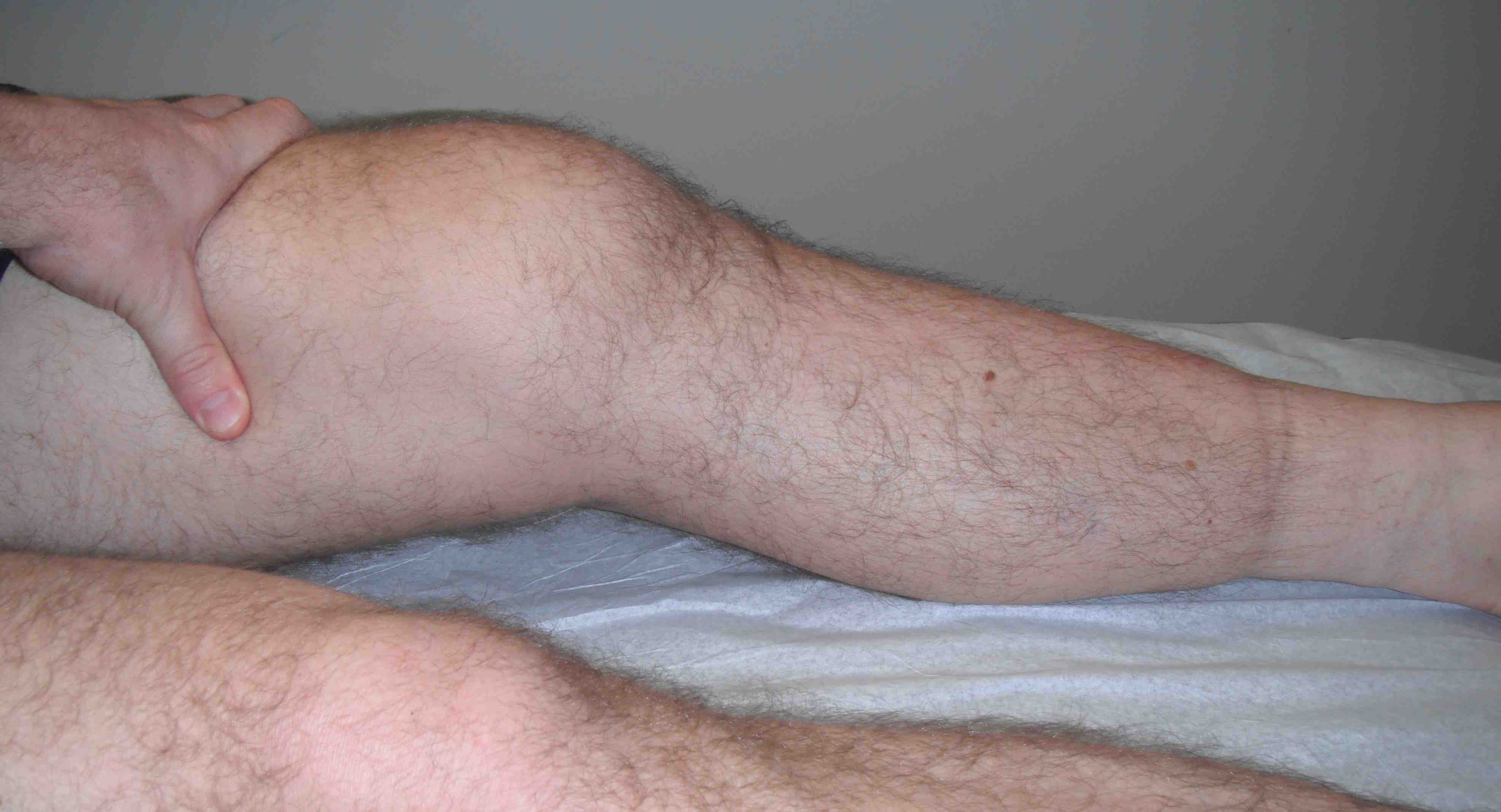
Effusion
- swipe, ballot, tap
Range
- FFD / Recurvatum / lift foot in air
- active extension / quads lag
- range of flexion bilaterally
FFD
- effusion
- entrapped meniscus
- ACL stump
- loose body
Feel
Flat
- Extensor mechanism
- patella
- tibial tuberosity
Flexed
- Joint lines, MCL, LCL
- tibial and femoral condyles
- popliteal fossa
Palpate distal femur for osteochondromas
Examine Ligaments
Collaterals
Test at 0 and 30o
- if loose at 0, loss of secondary stabilisers
Grading
1+ Surfaces separate 5mm or less
2+ 5 - 10 mm
3+ 10 mm or more
ACL / PCL
Lachmann's
- 85% sensitive awake
- 100% asleep
Check loss of tibial step off
- posterior sag
- MTP normally 1 cm anterior to MFC
Quadriceps active
- knee at 90o
- stabilise foot & ask to slide foot down bed
- N < 1mm / PCL > 3mm
Anterior / Posterior drawer
- restore tibial step off
Posterolateral drawer
- 30o IR
- tightens PLC
Posteromedial drawer
- 15o ER
- tightens PMC
Pivot Shift
- valgus stress with IR + axial compression
- knee moved from extension to flexion
- in chronic ACL deficiency, the LTC is subluxed anteriorly
- at 30o it reduces backwards
- this is when ITB passes behind axis of rotation and becomes flexor
- grade pivot glide / 1 / 2 / 3
Must have 4 things
- MCL to pivot about
- intact ITB
- no FFD
- ability to glide i.e. no meniscal pathology
PCL / Posterolateral Corner (PLC)
External rotation / Recurvatum
- hold big toe and assess PLC
- knee moves into recurvatum, tibia externally rotates & subtle varus
- indicates PCL + PLC + LCL
Reverse pivot shift
- with valgus and ER
- flexion to extension
- in flexion, the LTP is posteriorly subluxed
- ITB become extensor
- reduces as extend
- must compare with other side
- present in 30% normal population especially ligamentous lax
Dial test / Prone
- measure thigh foot angle
- examiner holds knees together
- increase at 30o only - PLC
- increases at 30 then again at 90 - PLC + PCL
- isolated PCL - no increase
- >10o compared with normal side
Meniscus
McMurray
- Flexion to extension
- Full IR - LM
- Full ER - MM
- i.e. test meniscus heel is pointing towards
- positive test is palpable / audible thud, snap, click
Squat test
- feet IR and ER
4Cs
Concealed / popliteal fossa
Cephalad / Hip
- rotation in flexion
- adduction / abduction in extension
Circulation
Collagen