Look
Aids
Shoes - raises / wear patterns
Stigmata generalised disease
Hands - RA, CMT
Front
Knee alignment
Forefoot - Hallux & Lesser toes
Scars
Circulatory changes
Medial Side
Turn affected side away & ask to step foot forward
Flexed attitude of knee
Medial arch - planus / cavus
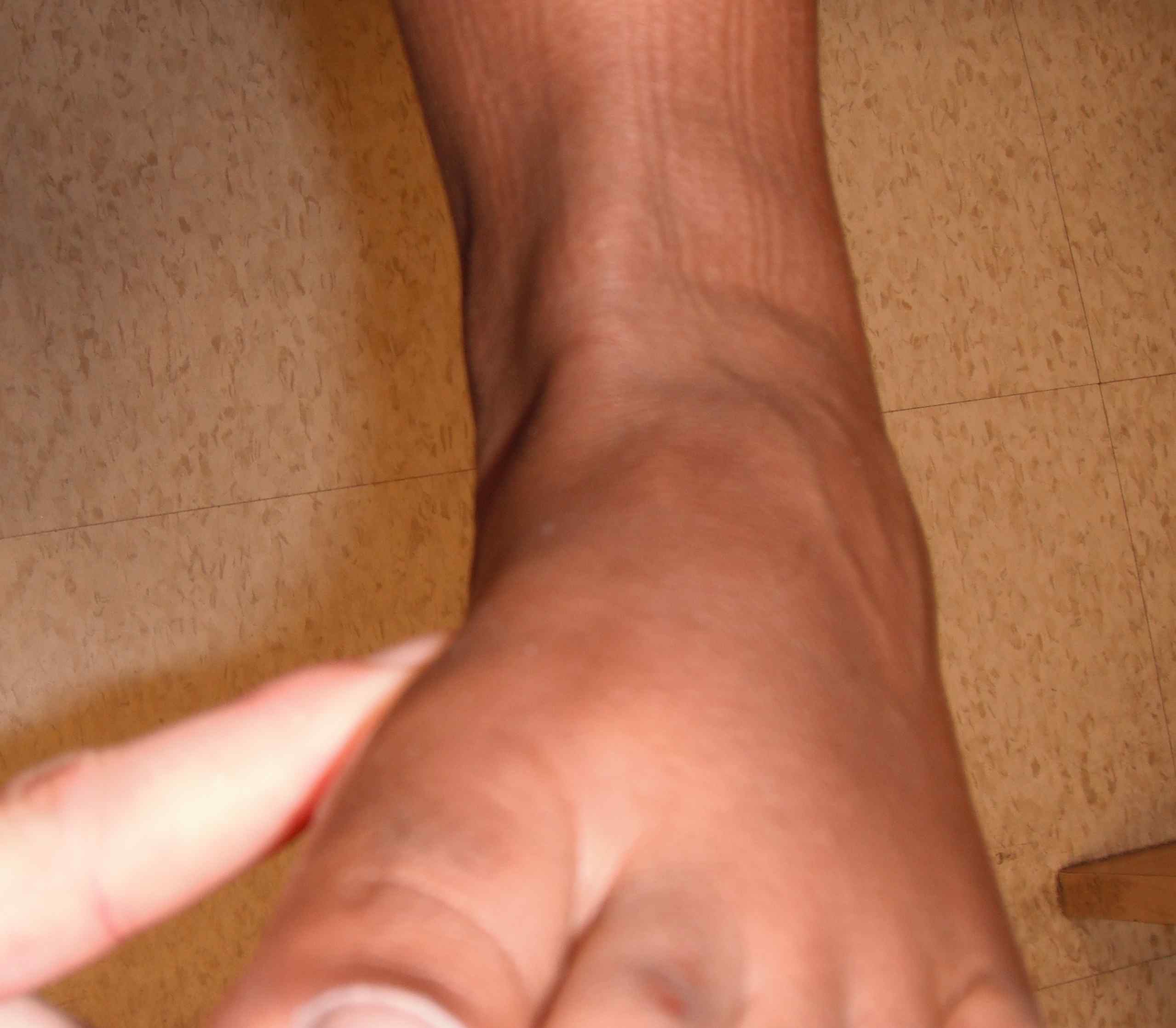
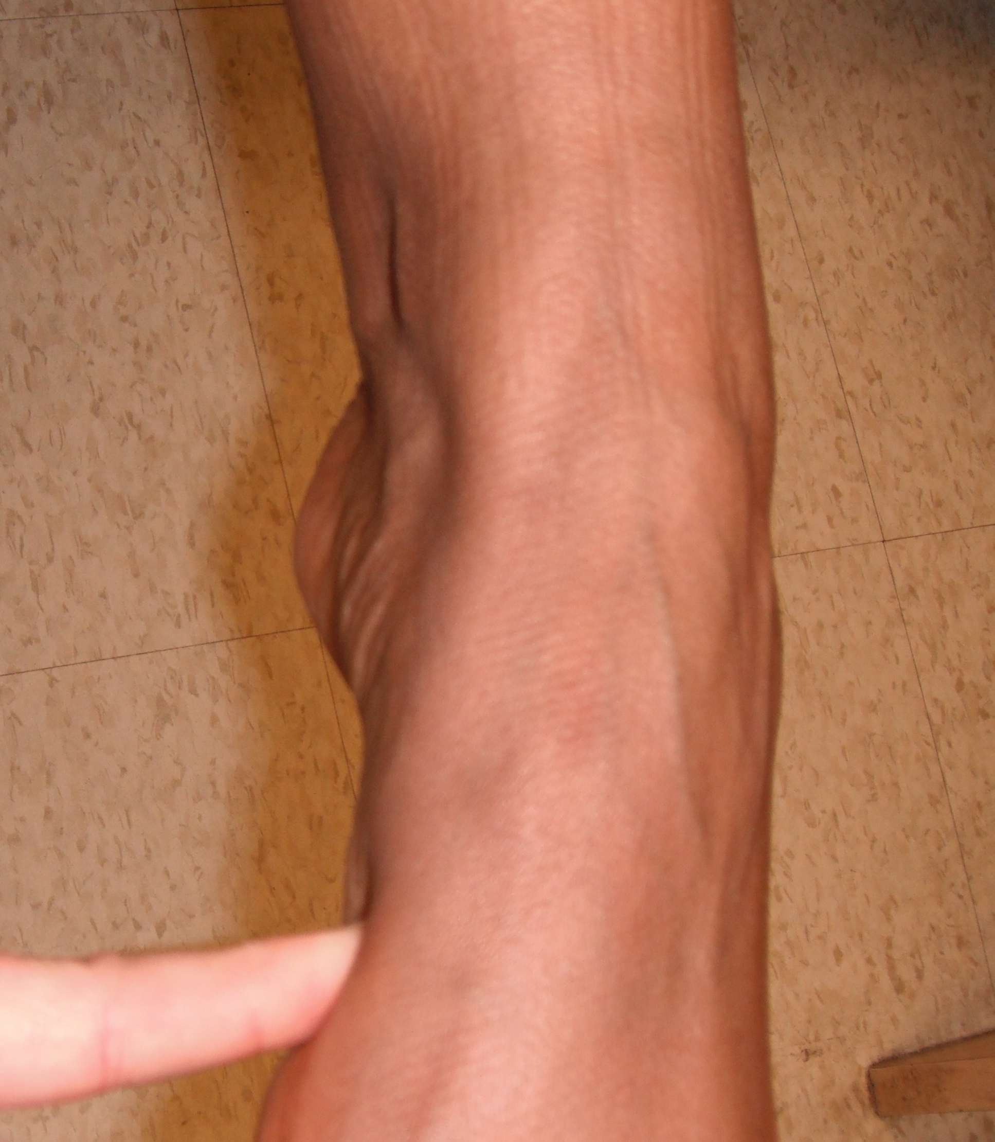
Behind
Spine - scoliosis / spinal dysraphism
Hindfoot varus / valgus
Forefoot (Too many toes)
Scars
Calf wasting
Double heel raise
- Heel swings into varus or remains in valgus
- ? mobile subtalar joint
- ? Medial arch restoration
Single heel raise
- Must put patient close to blank wall half a foot length from the wall
- otherwise will cheat by pushing up or leaning forward against wall
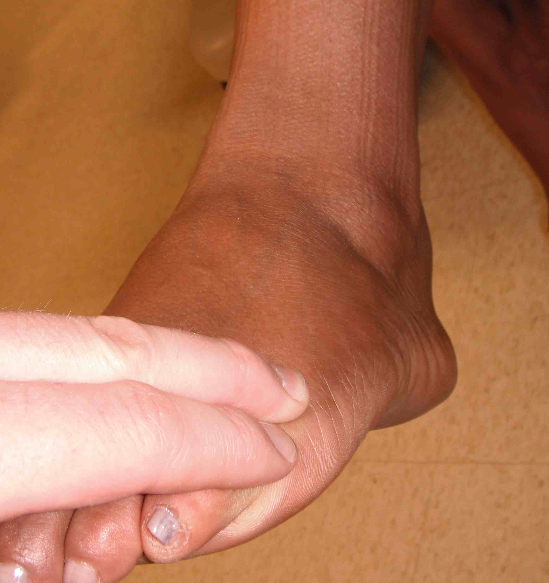
Lateral side
Haglund's
Peroneal tendons
Gait
Ankle
- Stiff / Fixed equinus
- Weak / Foot drop
- Painful / Antalgic
Foot Progression angle
Tip toe - strong S1
Heel walk - strong L4
Sit
On edge of bed with legs hanging
- examiner sits on chair
Screen active ROM AKJ and STJ bilaterally
Look at sole
- normal distribution weight pattern
- callosities
- lumps / plantar fibromatosis
Feel
" Where is it painful?"
Lateral aspect
- lateral malleolus
- lateral ligament complex
- Peroneal tendons
- sinus tarsi
- base of 5th
Posterior aspect
- tendo-achilles
- insertional / non insertional
Medially
- deltoid ligament
- tibialis posterior
- MT joints
- sustenaculum tali
Anterior
- ankle joint tenderness / effusion
- AITLF
Sole
- fat pad
- insertion of plantar fascia
Midfoot
Forefoot
- hallux rigidus
- sesamoids
- metatarsalgia
- Mulder test / interdigital tenderness
Move
DF - active and passive range 20o
PF - active and passive range 50o
Subtalar joint motion
- ankle in 90° DF
- thumb on talar neck to detect talar movement
- opposite hand cups heel and inverts & everts
- Inversion 10-15o
- Eversion 0-5o
Midtarsal joint
- Foot at 90 to lock ankle mortise
- Adduct foot 20o
- abduct foot 10o
- dorsiflex
- plantarflex
T Ach
- Tenderness along tendon /Insertion, ? lump
- test power / pain
Tibialis posterior
- Prominent with plantarflexion and inversion
- Thickening
- Tenderness
- Check power if abnormal
Peroneus brevis & longus
- thickening
- Tenderness
- Dislocation (resisted eversion)
- active eversion
Tibialis anterior
- Prominent with dorsiflexion and inversion
- Insertion
EHL & EDL - Dorsiflex toes
Special tests
Instability
Anterior drawer
- Due to complete tear of ATFL
- Grasp lower tibia and cup calcaneum
- "clunk" or draw
- compare with other side > 3 mm
Lateral instability
- Inversion stress
- Gaping of soft tissues
- Talar tilt (may occur in normal & must compare with other side)
- Needs to be confirmed on stress views
- > 20o
Medial instability
- Eversion stress
- Gaping / widening
- Needs to be confirmed on stress views
Gastrocnemius /soleus contracture
Test if limited dorsiflexion
Silverskiold Test
- Extend knee - dorsiflexion limited by both soleus & gastrocnemius contracture
- Flex knee - gastrocnemius relaxed (crosses knee joint)
- If dorsiflexion still limited it is due to soleus contracture
- If limited in extension & not in flexion then due to gastrocnemius contraction
Pes cavus
Claw toes - flexible / fixed
Individual power compared with other side
- Tibialis anterior (inversion in DF)
- Tibialis posterior (inversion in PF)
- Peronei
Coleman block test
- Dynamic visualisation of hindfoot correction
- Stand on 2cm block
Passive correction of plantar-flexed 1st MT
Spine / Neuro Exam
Hallux valgus
MTPJ Painful / limited range
- flexion 45o
- extension 70-90o
- redo range with correction
IPJ
- hallux interphalangeus
- extension / flexion
Lesser toes
- fixed / mobile
- dislocated
Concealed
Spine
Neuro exam
Vascular exam
Ligamentous laxity