Background
Deformities
Mallet
- DIP flexed
- MTP / PIPJ neutral
Hammer
- PIP flexion
- DIPJ neutral / extended
Simple - MTP not involved
Complex - MTP hyper-extended
Claw
- PIPJ and DIPJ flexed
- MTPJ hyperextended
Curly toe
- PIP and DIP flexion
Mallet
- DIP flexed
- MTP / PIPJ neutral
Hammer
- PIP flexion
- DIPJ neutral / extended
Simple - MTP not involved
Complex - MTP hyper-extended
Claw
- PIPJ and DIPJ flexed
- MTPJ hyperextended
Curly toe
- PIP and DIP flexion
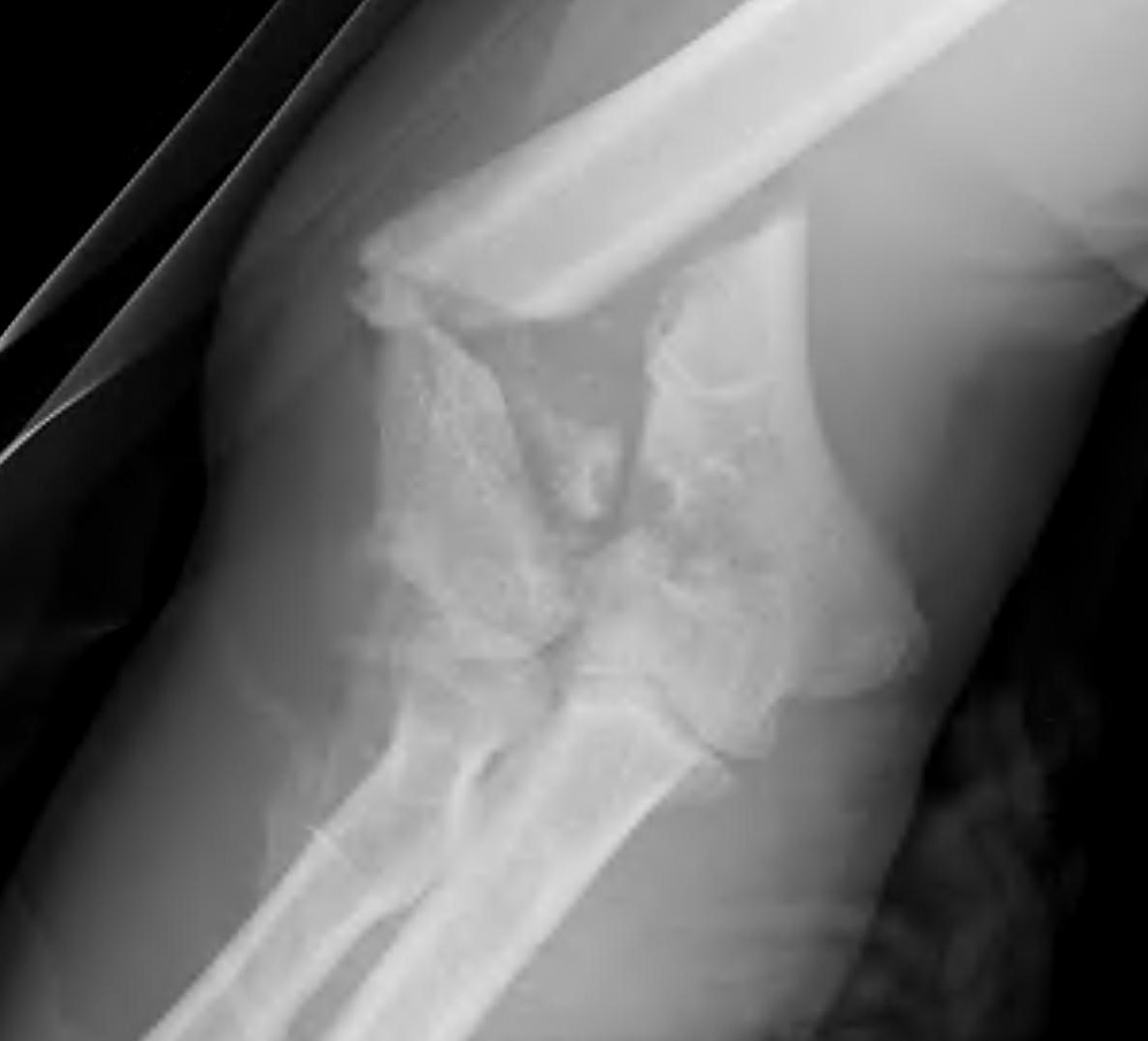
1. TFCC
- central articular disc
- TFCC is major stabiliser of DRUJ
- arises ulnar aspect of lunate fossa of radius
- inserts fovea at base of ulna styloid
2. Dorsal and Palmar Radio-Ulna ligaments
Thick fibrous structures
- from ulna styloid
- important stabilises of DRUJ
In normal wrist
- Dorsal RU ligament tight in pronation
Nerve supply
- C7, C8, T1
- nerve picks up some branches of C7 from the lateral cord
Origin
- direct continuation medial cord
- runs between the brachial artery and the vein in the arm
- behind MCNFA
Arm
- pierces the medial intermuscular septum to run in posterior compartment
- runs anterior to the triceps
Elbow
Traumatic initial cause in 95%
M:F 2:1
Age of initial dislocation inversely related to recurrence rate
- patients younger than 20 have a redislocation rate of 90%
- between 20 - 40 years, redislocation rate of 60%
- patients > 40 years have a 10% rate of dislocation but a higher rate of cuff tears (up to 40% in patients > 60yrs)
Synovial joint with hyaline cartilage
Has fibrocartilage intra-articular disc
- complete or incomplete
- usually degeneration by 4th decade
Clavicle may lie superior to acromion in normal population
Acromioclavicular Ligaments
I DIPJ
II Middle Phalanx
III PIPJ
IV Proximal Phalanx
V MCPJ
VI Metacarpal
VII Dorsal Wrist Retinaculum
Injury to ulnar collateral ligament of thumb MCPJ
Initial description
- chronic laxity of British gamekeeper's thumb's
- no specific trauma
- secondary to breaking pheasant's neck
Acute trauma
- snow ski
- ball games
Valgus / forced abduction
UCL
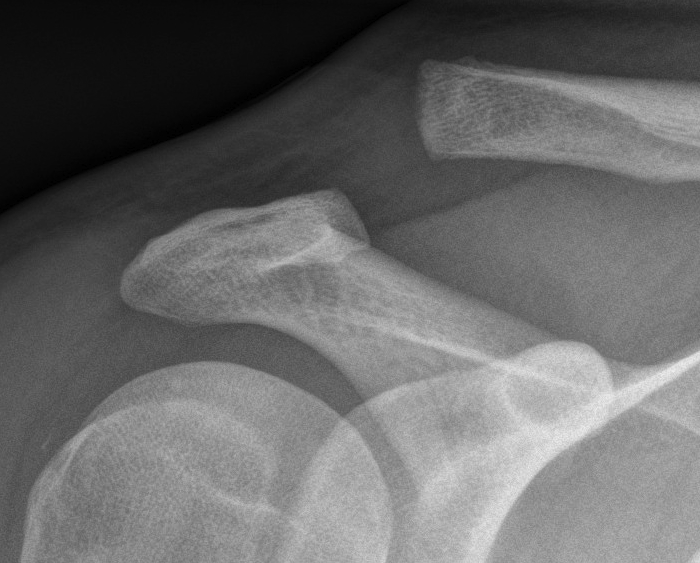
Intra-articular proximal ulna fracture
Articulates with trochlea
- may have a central bare area
Triceps insertion
- via broad aponeurosis which blends with anconeus and CEO
Undisplaced fracture
- need to ensure triceps mechanism is intact
2 groups
- young patient with high velocity injury
- older patient with comminuted, osteoporotic fracture
In the second group fixation can be very difficult
Hinged Joint
- trochlea axis is centre of rotation
- 40o anterior angulation in sagittal plane
Lateral : Medial 9:1
4th & 5th decades
- M = F
- 75% dominant arm
50% of regular tennis players
- especially > 2 hrs / week
Insertion pathology / Enthesopathy
Over-extension of the elbow with supination / pronation
Lateral epicondyle
- anconeus from posterior face
- ECRB and EDC from anterior face (CEO)