Definition
Avascular necrosis of the immature femoral capital epiphysis
- self limiting
- goes through defined stages over 2-5 years
- can cause femoral head deformity and lead to osteoarthritis
Epidemiology
1/10,000 in Caucasians
- age 4 - 8
- male 4 x females
Bilateral - between 8 - 24%
Etiology
Lateral epiphyseal artery occlusion
Theories of cause
| Genetic | Vascular | Coagulation disorder | Environmental |
|---|---|---|---|
|
Unclear if this is true in twin studies
Likely enviromental |
Increased intra-articular pressure
Increased intra-osseous pressure |
Factor C / S deficiencies |
Lower socioeconomic
Short stature / low birth weight
Malnutrition
Parental smoking smoking
|
Transient synovitis
Xinling et al J Orthop Surg Res 2024
- systematic review of prevalence of Perthes in transient synovitis
- overall prevalence Perthes 3%
- recurrent transient synovitis: prevalence Perthes 36%
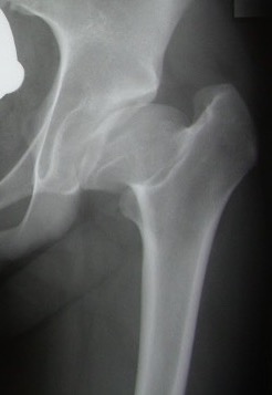
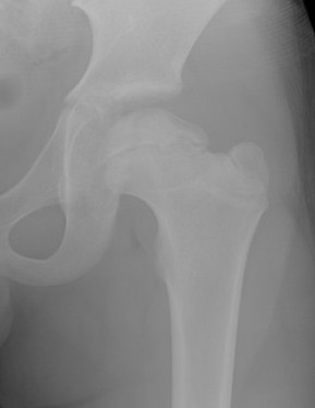
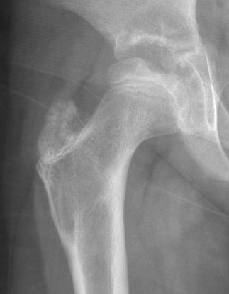
Pathology
Physis
- loses height
- increases width
- focal lateral collapse under acetabular margin
Coxa magna and partial subluxed head
Coxa breva
- physeal growth arrest
- prominent greater trochanter
Classification
Waldenstrom - natural history / chronological
Herring / Catterall - prognosis / treatment based upon extent of head involvement
Stuhlberg - long term outcome based upon residual femoral head deformity
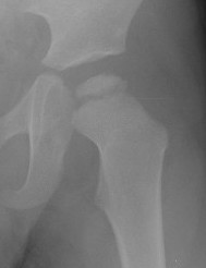
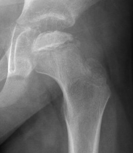
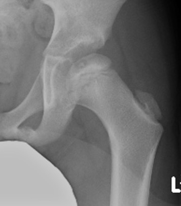
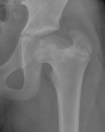
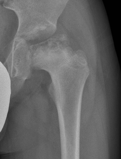
Waldenstrom classification
| Stage I | Stage II | Stage III | Stage IV |
|---|---|---|---|
| Necrosis | Fragmentation | Reossification | Remodelling |
| Dense flattened epiphysis | Resorption of necrotic bone | New bone formation | Head larger / neck shorter |
| 6 - 12 months | 12 months | 2 - 3 years | Until skeletal maturity |
 |
|
 |
 |
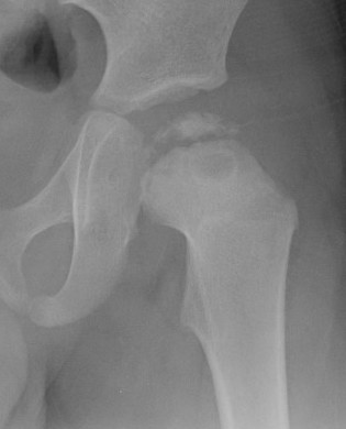
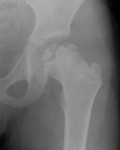
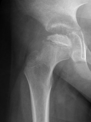
Herring lateral pillar classification
AP xray when disease in fragmentation
- divide femoral head into 3 pillars
- lateral (25%) / medial (25%) / central (50%)
- classification based upon lateral pillar involvement
- lateral pillar involvement leads to subluxation and deformity
- added group B/C: 50% involvement
| Group A | Group B | Group C |
|---|---|---|
| No lateral pillar involvement | > 50% lateral pillar intact | < 50% lateral pillar intact |
 |
 |
|
Catterall classification
Based upon extent of involvement of femoral head
- Grade I: < 25%
- Grade II: < 50%
- Grade III: < 75%
- Grade IV: Entire femoral head
Stuhlberg classification
Estimate long term prognosis of the hip based upon final joint morphology
- increasing deformity
- increasing risk of developing early osteoarthritis
- modified into 3 groups
| Group A | Group B | Group C |
|---|---|---|
|
Class I / II |
Class III
|
Class III/IV |
|
Round femoral head |
Ovoid femoral head |
Flat femoral head |
|
Low risk OA |
Moderate risk OA |
High risk OA
|
 |
 |
 |
- 58 hips followed for 20 years
- incidence OA: spherical hips 22%, ovoid hips 61%, flat hips 62%
- 88 hips followed for 21 years
- incidence OA: spherical hips 3%, ovoid hips 15%, flat hips 54%
Prognosis
Poor prognostic factors
- age > 8 at age of diagnosis
- females do worse / less time for femoral head remodel
- lateral pillar collapse / Herring C
- extent of head involvement / Catterall
- lateral extrusion of femoral head
- poor range of motion
- 345 Perthes hips
- worse outcomes age onset > 8 / females / Herring group C
Clinical
Hip / knee pain
Short stature
Antalgic / Trendelenberg gait
Decreased ROM
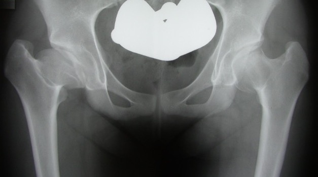
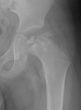
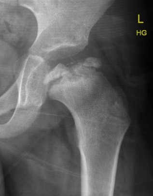
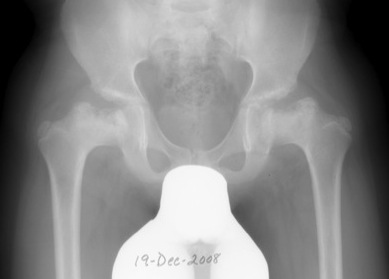
Xray
Lateral pillar involvement guides management
No lateral pillar involvement versus significant lateral pillar involvement
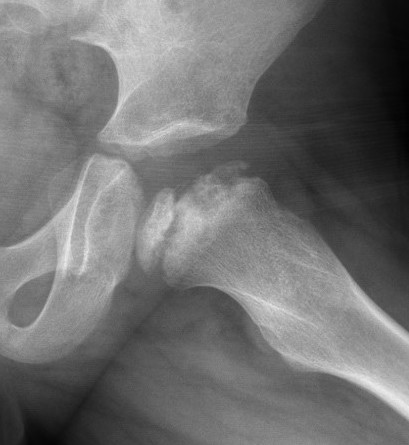
Containment - if the head is not contained within acetabulum, guides management
Contained versus uncontained hip
Hinge abduction
Abducted hip does not obtain full coverage of femoral cartilage / full containment
- hinging on portion of femoral head
- varising femoral osteotomy will worsen symptoms
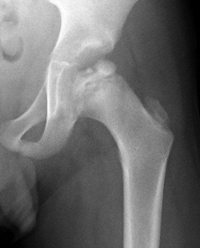
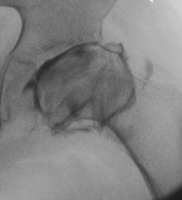
Intra-operative arthrogram
Findings
- hinge abduction / rose thorn appearance
- medial pooling
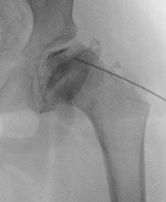
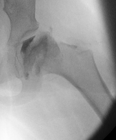
Hinge abduction with rose thorn appearance
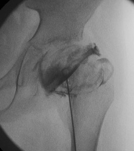
Hinge abduction with rose thorn appearance
MRI
Uses
- early diagnosis of Perthes / MRA
- assess containment / amount of cartilaginous head outside of acetabulum
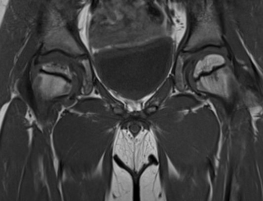
Central Perthes on MRI
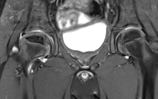
Early presentation of Perthes with contained hip
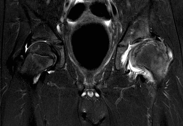
Uncontained left hip with Perthes
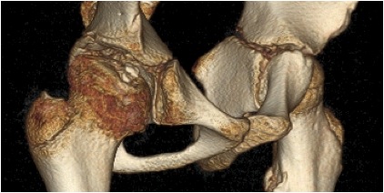
CT
Differential diagnosis
AVN
MED / SED
Hypothyroidism
Multiple epithelial dysplasia
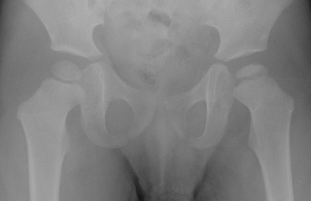
- bilateral and symmetrical
- acetabular involvement
- no metaphyseal cysts
- other joint involvement
- consider skeletal survey in those with bilateral "perthes"
Multiple epithelial dysplasia with bilateral symmetrical involvement of both hips