

Incidence
3% of knee injuries
2 Groups of patients
1. Patients with no predisposition to patella instability
- traumatic injury
- direct lateral blow to patella / twisting injury
2. Patients with anatomic predisposition to instability
- atraumatic / minimal trauma
- young / valgus malalignment / ligamentous laxity / malrotation
Medial Patellofemoral Ligament (MPFL)


Anatomy
Second layer of the knee / deep to retinaculum / superficial to capsule
Origin
- medial femoral condyle
- between medial femoral epicondyle and adductor tubercle
- superior to origin of MCL
Insertion
- superomedial patella
- broad insertion 2 - 3 cm
MPFL tears with patella dislocation
- systematic review of MPFL injury after 2500 patella dislocations
- incidence MPFL injury 95%
- injury location: patella 37%, femur 37%, combined 25%, midsubstance 15%
Osteochondral fractures
Clinical
Associated with hemarthrosis after patella dislocation
Isolated MPFL tear does not cause hemarthrosis

Large hemarthrosis left knee
Incidence
- systematic review of 3000 patella dislocations
- first time dislocation: 43%
Location
Uimonen et al OSJM 2021
- 134 patients with osteochondral fracture after patella dislocation
- patella 63%
- lateral femoral condyle 34%
- both 3%
- systematic review of 3300 patella dislocations
- overall prevalence of osteochondral injury 49%
- medial patella most common site
- location: medial patella 37%, central patella 24%, LFC 21%, lateral patella 12%
Xray
Look for osteochondral fractures
- skyline xray: suprapatella pouch
- lateral xray: notch
- AP xray: gutters

Osteochondral fracture visible on skyline view


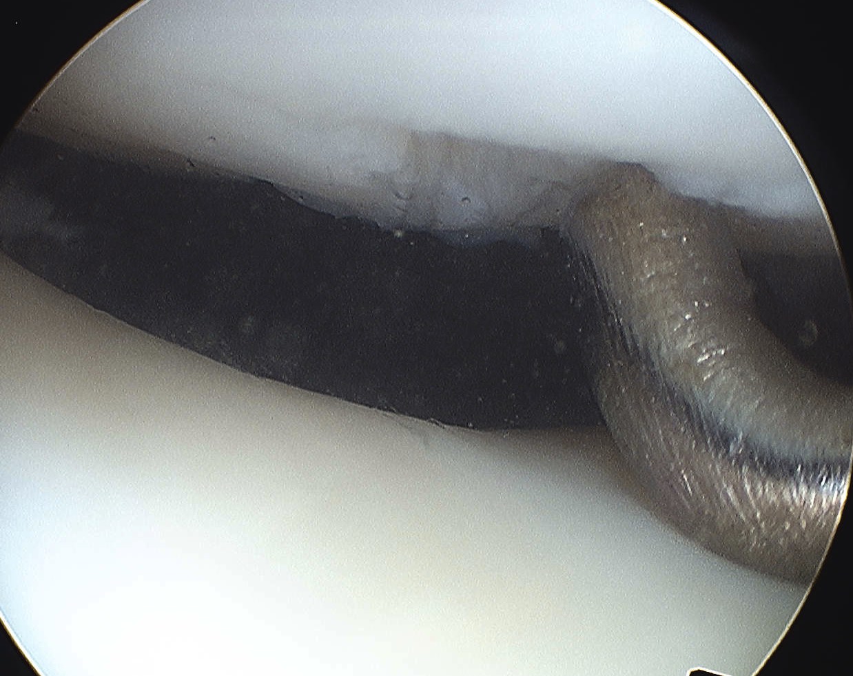
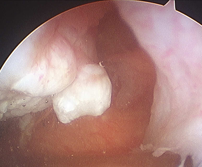
Loose body in notch with donor site from patella


Loose body in lateral gutter


Large medial patella osteochondral fracture


Large lateral femoral condyle osteochondral fracture
CT


Osteochondral fracture of the lateral femoral condyle


Large osteochondral fracture medial facet patella


Large osteochondral fracture lateral femoral condyle
MRI
Advantage
- identify smaller chondral lesions
- identify size of osteochondral fragment better than CT
- diagnose MPFL tear location
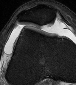
Small chondral fracture in notch from medial facet patella, and avulsion of MPFL from patella


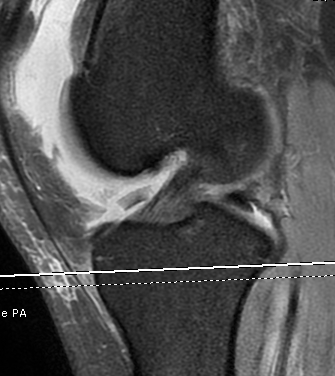
Large chondral fracture from medial facet of patella


Large chondral fracture from medial facet of patella


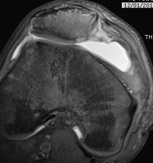
Osteochondral fracture lateral femoral condyle
Management
Issues
1. Osteochondral fracture
- removal versus ORIF
- depends on size
2. Operative management of first time patella dislocation
Osteochondral fracture
Options
< 1 cm2 - remove
> 1 cm2 - ORIF
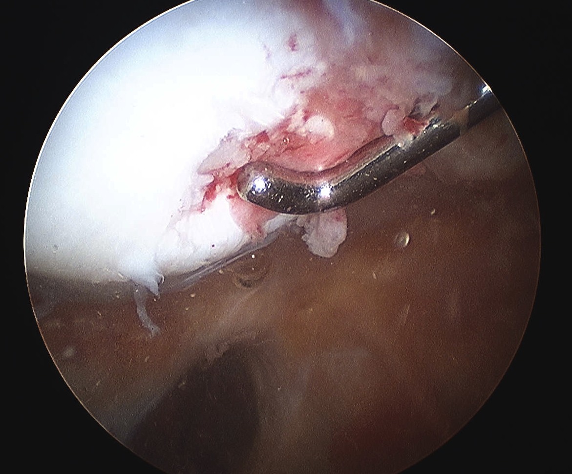
Arthroscopy and fragment removal
Small irreparable chondral fracture from central patella
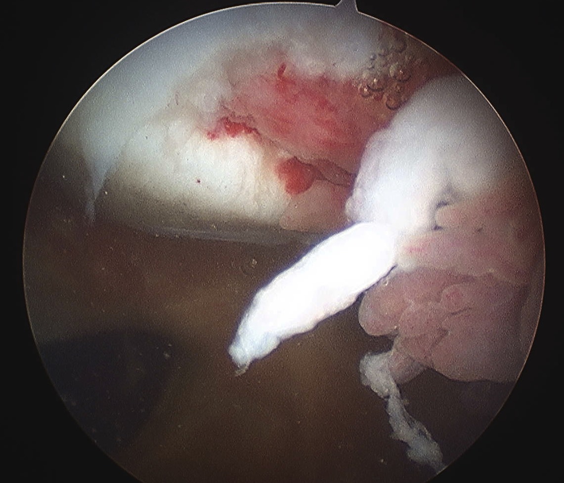
Small irreparable chondral fracture from central patella


Osteochondral fragment in notch from uncontained defect lateral femoral condyle
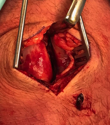
Open reduction and internal fixation
Approach
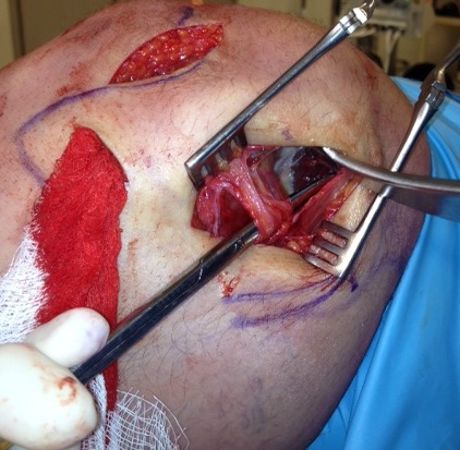
Patella - medial parapatellar approach with knee extended
Lateral femoral condyle - lateral parapatellar approach with knee flexed to 90
Fixation
Headless compression screws
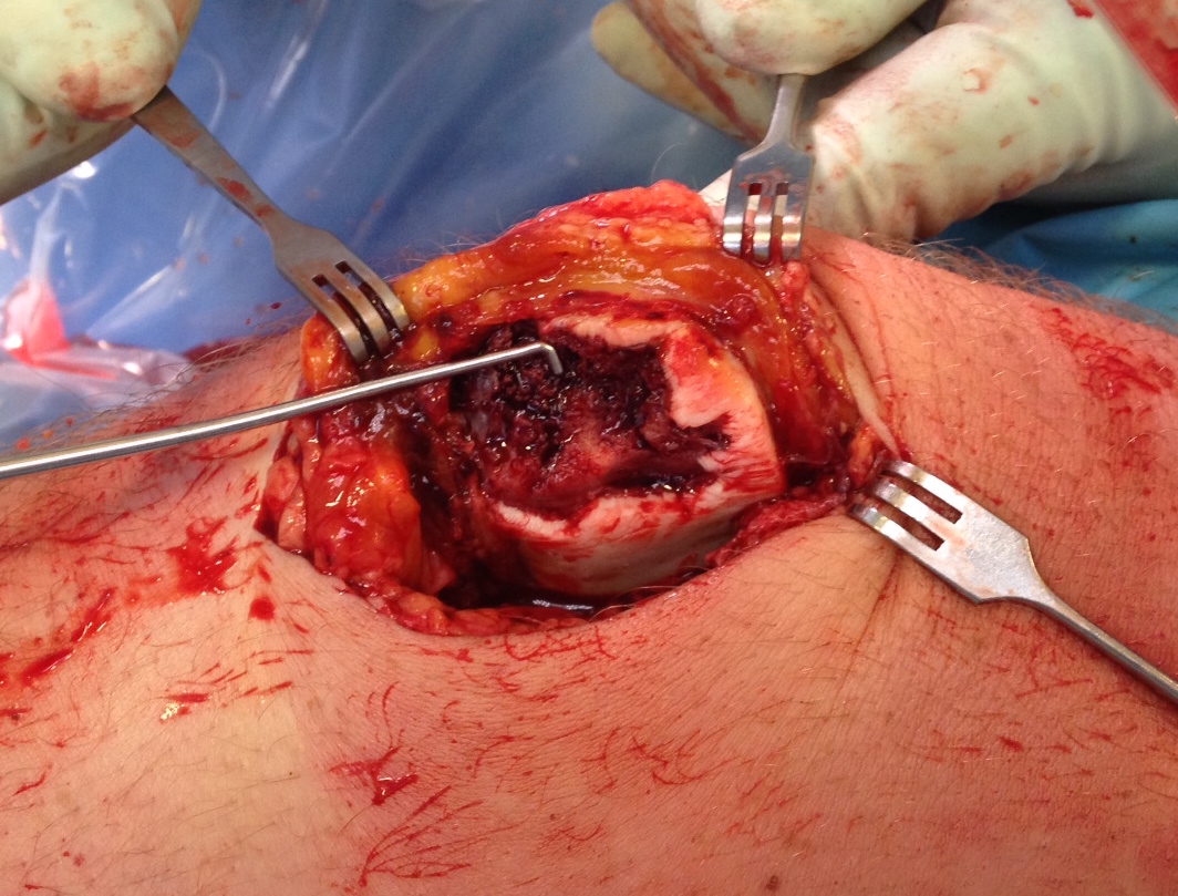
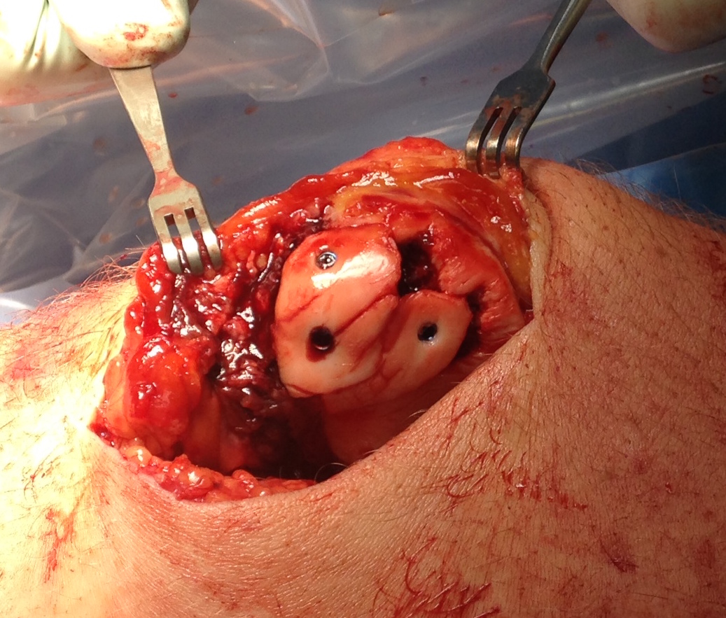
Large osteochondral fracture medial facet patella


Large osteochondral fracture medial facet patella


MPFL repair in first time patella dislocation
Indication
Patient undergoing surgery for osteochondral surgery
- International Patellofemoral Study group
- guidelines for management of first time PFJ dislocation
- no osteochondral fracture: non operative
- osteochondral fracture removal or repair: operative management patella instability
- most recommend reconstruction
Issues
MPFL repair
- need MRI to identify MPFL tear location and repair appropriately
- longer rehabilitation especially if fragment removal only
Estimate patient recurrence rate
- some patients low risk of recurrence / traumatic dislocation
- some patients very high risk of recurrence / need MPFL reconstruction not repair
Outcomes
Operative versus non operative
- meta-analysis of RCTs of operative v nonoperative in first time patella dislocation
- 306 patients, 56% female
- recurrence rates: operative 11% versus 30%
Longo et al Clin J Sports Med 2017
- systematic review of 2000 first time patella dislocation
- recurrent instability nonoperative: 36%
- recurrent instability operative: 25%
Adolescents / high risk recurrence
- 41 adolescents undergoing surgery for osteochondral fracture after first time dislocation
- 61% recurrent patella instability
- TTTG > 15 mm: recurrent instability 75%
- TTTG > 20 mm: recurrent instability 86%
- MPFL repair did not alter instability
MPFL repair versus reconstruction
- meta-analysis of RCTs of operative v nonoperative in first time patella dislocation
- recurrence rates: MPFL repair 16%, MPFL reconstruction 4%