xrays
Metacarpal Fractures
Fractures
1. Neck of 5th Metacarpal
2. Metacarpal Shaft
3. Metacarpal Head
4. Base of Metacarpal Fracture Dislocations
5. Base of Thumb Fractures / Bennett's / Rolanda
1. Neck of 5th Metacarpal Fracture
Non operative Management
Accept 45o angulation
- will have finger extensor lag, but will recover
Ulna collateral ligament injury
Aetiology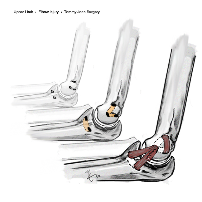
Throwing injury
- seen in the throwing athlete
- repetitive microtrauma / valgus stress
- develop laxity
History
Initially
- lose velocity / accuracy
Develop medial pain
40% ulna nerve symptoms
Background
Aetiology
Intrinsic
- inflammatory
- degenerative
Extrinsic
- traumatic
- spur
Epidemiology
F > 40
Associations 60% of cases
- hypertension
- diabetes
- obese
- trauma
- prior surgery
- steroids
Ankle Arthroplasty
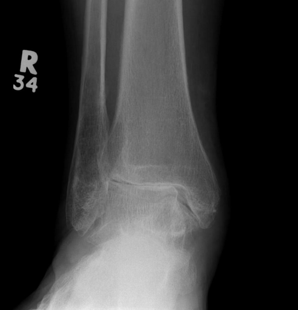
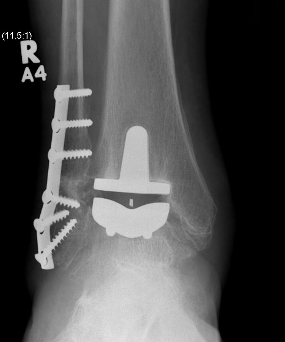
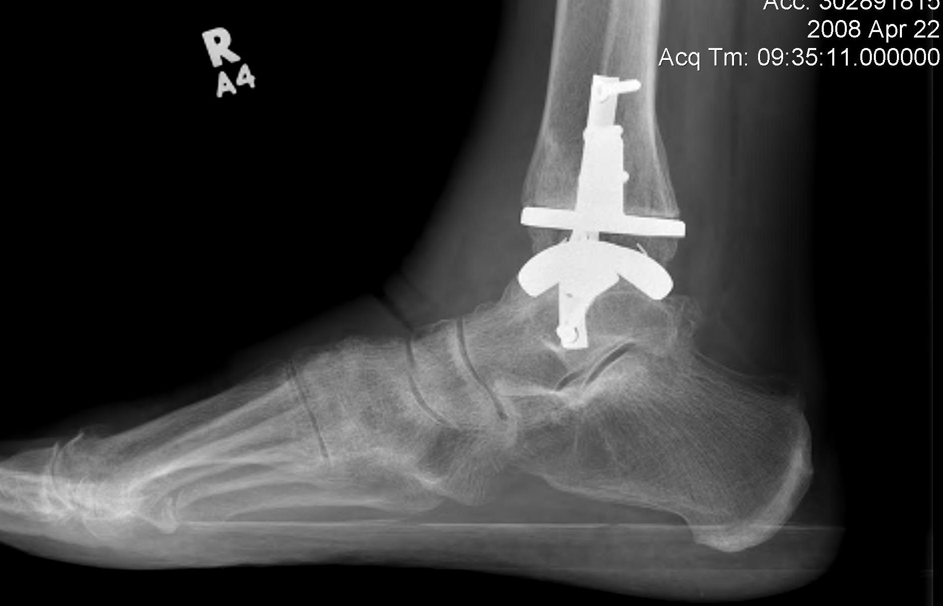
History
First generation (late 70s early 80s)
Results
Describing Bone Tumour X-rays
1. Pattern of bone destruction
Geographic
Least aggressive
- usually indicative of slow growing lesion
- usually seen in benign tumours
- may be myeloma / mets / OM
Narrow transition from normal to abnormal bone
- Margin of the lesion is well defined
- margin is easily separated from surrounding bone
- margin may be smooth / irregular, sclerotic / non sclerotic
Kienbock's disease
Definition
Avascular necrosis & subsequent disintegration of lunate
Aetiology
50-75% history of trauma
Occasionally seen in sickle cell / steroid use
Pathogenesis
Vascular Theory
Trauma disrupting vascularity
- single incident with disruption of blood supply
Olecranon fractures
Definition
Intra-articular proximal ulna fracture
Anatomy
Articulates with trochlea
- may have a central bare area
Triceps insertion
- via broad aponeurosis which blends with anconeus and CEO
Management
Non operative Management
Undisplaced fracture
- need to ensure triceps mechanism is intact
Gout
Definition
Heterogeneous group of diseases characterised by
- hyperuricaemia
- recurrent attacks of acute arthritis
Diagnosis confirmed by
- crystals of Monosodium Urate in synovial fluid
- tophi ("Porous stone") urate in soft tissues
- renal urate stones
Epidemiology
Adult men