techniques
Deltoid ligament injury


Etiology
Ankle sprain
- eversion / external rotation
Ankle fractures
Radiocarpal fracture dislocations
A. Radiocarpal Dislocation


Distal humeral physeal separation
Pathology
Children < 6
- entire distal humerus physis is displaced
Xray
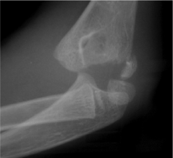
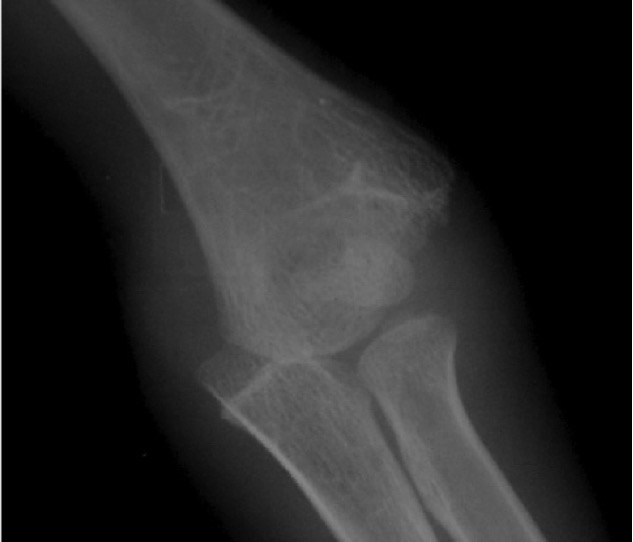
Distal physis not ossified < 1 year
- may be a difficult diagnosis
Trochanteric Osteotomy
Types
1. Standard trochanteric osteotomy
2. Sliding trochanteric osteotomy
3. Extended trochanteric osteotomy
Standard Trochanteric osteotomy


Tibial tubercle fractures
Epidemiology
Adolescent boys
Ossification
Proximal tibia / primary ossification centre
Tibial tuberosity / secondary ossification centre
- eventually merges with primary ossification centre
Ogden Classification
Type I - Tibial tuberosity ossification only
ACL rupture pediatric
Issues
1. More common recently
- more high level sport
2. High risk of reinjuring knee from instability
- can suffer permanent severe chondral and meniscal damage
3. Risk of physeal arrest high if bone block across physis
- risk is growth arrest with ACL reconstruction
Epidemiology
Hemarthrosis
- 60% children have ACL tear
TFCC tears
Definition
Present with pain but not instability
Types
Traumatic
Degenerative
Different treatment algorithms for each
History
Ulna side wrist pain
- may be worse with rotation
- opening doors and jars
History of trauma
Examination
Local tenderness DRUJ
Stems
Advantage
1. Reduce implant loosening
- offset load sharing to diaphysis
- 30% if > 70 mm
2. Restore optimal alignment
Indications
1. Using augments or bone grafting
2. Increased constraint
- VVS / hinge