ossification
Calcification v Ossification
Xray
Calcification
- central pattern
- often increased opacity compared with bone
Ossification
- peripheral pattern
- similar density to bone
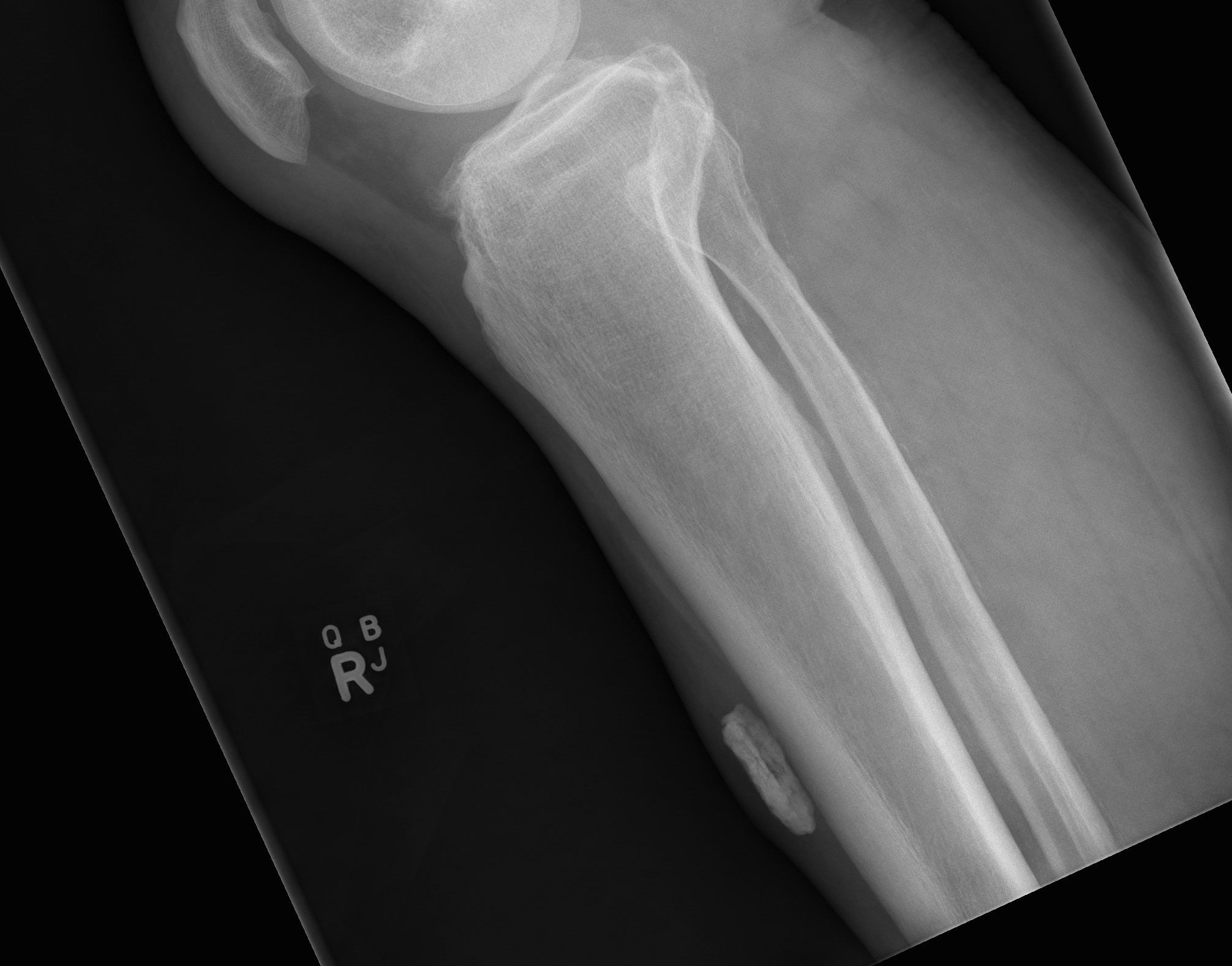
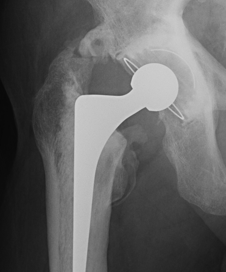
Centers of ossification
Age of appearance in girls
C (capitellum) 1
R (radiua) 3
I (internal epicondyle) 5
T (trochlea) 7
Pelvic and acetabular fractures
Anatomy
Bones more elastic and malleable
- absorb much more energy
Very thick periosteum
- can be periosteal sleeve fracture
Ossification
Triradiate cartilage fuses 13-16
Iliac / Ilium / ASIS apophysis
- appear as teenager
- fuse a couple of years later
- can confuse with fracture
Classification Key & Conwell 1951
Congenital odontoid abnormalities
Ossification
Atlas
Three 1° Ossification centres
- two lateral masses with posterior arch in between
- anterior arch which appears at 1 year
- posterior arch fuses at 4 years
- anterior & posterior fuses to body at 7 years
Axis
Five 1° Ossification centres
- two neural arches / lateral masses
- two 1/2 dens
- body
Five 2° Centres