Approaches
Posterolateral
Anterior
Anterolateral
Posterior
Medial
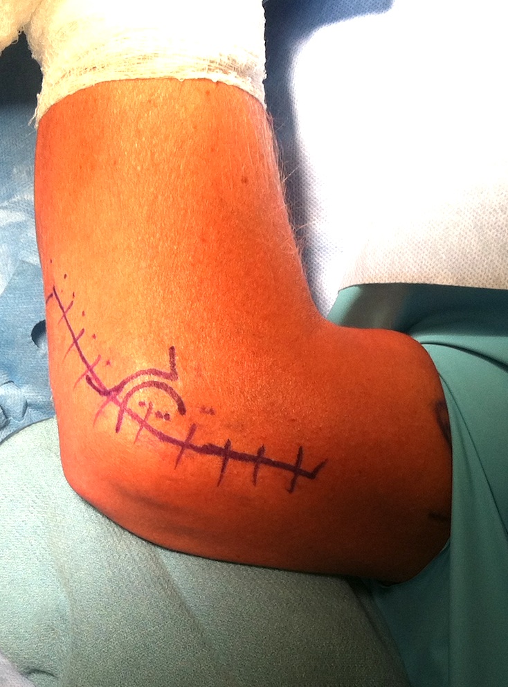
Posterolateral / Kocher
Concept
- between ECU and anconeus
Indications
- radial head ORIF / replacement
- washout elbow joint
Technique
Position
- patient supine with arm on hand table
Landmarks
- lateral epicondyle, head radius, olecranon
Incision
- proximally over lateral supracondylar ridge 5cm proximal to elbow
- continue 5cm distal towards radial head
- curve posteriorly to ulna border
Inter-nervous plane
- between ECU (PIN) & anconeus (RN)
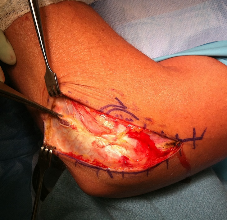
Superficial dissection
- identify the plane between the anconeus & ECU
- anconeus triangular muscle fanning from lateral epicondyle out to olecranon
- interval best identified distal to epicondyle
Deep dissection
- fully pronate the forearm to move the PIN away
- elevate ECU and EDC off capsule anteriorly
- keep incision anterior to avoid dividing lateral ulna collateral of LCL
- LCL in line and deep to anterior fibres of anconeus
- divide capsule over radial head
- do not continue below the annular ligament or retract too vigorously to avoid damage to the PIN
Extension
- proximally between triceps and BR/ECRL anteriorly
Anterior Approach
Concept
- between Biceps and BR proximally
- between BR and PT Distally
Indications
- repair of median nerve / radial nerve / brachial artery
- reinsertion of biceps tendon
Technique
Incision
- S shaped incision over the anterior aspect of elbow
- 5cm above the flexion crease on medial side of biceps
- curve across the front of elbow joint
- continue laterally along medial aspect of BR
- don't cross flexion crease at 90o
Internervous Plane
- between the BR (radial nerve) and Brachialis (MCN) proximally
- between BR (radial nerve) and PT (median nerve) distally
Superficial Dissection
- incise deep fascia in line with skin incision and ligate veins
- lateral cutaneous nerve of forearm located and preserved
- lacertus fibrosis identified and cut at the origin with the biceps tendon
- brachial artery beneath lacertus
- median nerve lies medial to artery
- radial nerve found between the brachialis and BR
- passes lateral to biceps tendon
Deep dissection not required
Dangers
- lateral cutaneous nerve of forearm located between the Brachialis and Biceps
- brachial artery immediately deep to lacertus
Extensions
- proximally along the medial side of the biceps to expose the brachial artery
- distally as anterior / Henry approach to forearm
Anterolateral Approach
Concept
- between BR / radial nerve and biceps / PT
Indications
- ORIF of capitellar fractures
- OCD of capitellum
- tumors of the proximal radius
- PIN compression
- distal biceps rupture
Technique
Incision
- 5cm above the flexion crease of elbow over the lateral border of biceps muscle
- small curve at flexion crease of elbow
- extends distally following the medial border of brachioradialis
Internervous Plane
- proximally between BR (radial nerve) and Brachialis (MCN)
- distally between the BR (radial nerve) and PT (median N)
Superficial dissection
- preserve LCN of forearm (superficial to deep fascia in interval between biceps and brachialis)
- incise deep fascia along the medial aspect of BR
- identify and protect radial nerve proximally between the BR and brachialis
- brachialis / biceps reflected medially and BR reflected laterally
Deep dissection
- follow the radial nerve until divides into the SRN / PIN and motor branch to ECRB
- develop plane between BR and PT
- will have to ligate the recurrent vessels (leash of Henry) here that enters BR
- retract radial artery and PT medially
- divide capsule longitudinally between the radial nerve laterally and the brachialis medially
- the proximal radius is further exposed by fully supinating the forearm
- detaching the supinator from the oblique line to avoid damage to the PIN
Dangers
- PIN
- radial nerve
- recurrent branches of radial artery
- lateral cutaneous nerve of forearm
Extension
- proximally by conversion into anterolateral approach to the humerus
- distally extended as the anterior approach to the forearm
Posterior Approach
Indication
- ORIF distal 1/3 humerus
Technique
Position
- patient on side, arm over bolster
Incision
- midline and extending distally
- curve laterally about the tip of olecranon
- avoids sensitive scar
Superficial dissection
- identify ulnar nerve medially
- dissect from its bed (divide Osbourne's fascia) and vessiloop
Deep dissection
1. Mobilise medial and lateral sides of triceps
- beware radial nerve proximally on lateral side
2. Intra-articular fracture
- chevron osteotomy
- predrill and tap the olecranon for 6.5 mm screw
- Chevron osteotomy 2 cm from tip with osteoclasis of articular surface
- elevate the triceps superiorly off the humerus with olecranon
- can extend to lower 1/4 - any higher can endanger the radial nerve in groove
- cannot extend proximally but able to extend distally to expose the entire surface of the ulna
Medial Approach
Indications
- ORIF coronoid process fracture
- ORIF medial epicondyle
Technique
Incision
- curved incision on the medial aspect of the elbow 8-10 cm length
- centered on the medial epicondyle
Internervous Plane
- proximal - Brachialis (anterior) and Triceps (posterior)
- distal - PT and Brachialis
Superficial dissection
- locate the ulnar nerve and divide the fascia over the nerve
- mobilise and retract the ulna nerve posteriorly
- identify CFO
Options
1. Osteotomy medial epicondyle and reflect CFO
2. Open plane between PT and FCR
Dangers
- median nerve or AIN palsy with traction of the medial epicondyle
- ulnar nerve injury
Distal extension
- is limited by the median nerve