Definition
Rare congenital deformity causing rigid flatfoot
- rockerbottom foot
- fixed dorsal dislocation of talonavicular joint
Epidemiology
1/10,000
50% bilateral
Some autosomal dominant family history
50% associated with neuromuscular disorder / syndromes
- spina bifida / spinal muscular atrophy
- arthrogryposis
- neurofibromatosis
- Trisomy 15-18
Pathology
| Bony | Tendons | Joint / ligament |
|---|---|---|
|
Calcaneum - plantar flexed
Talus - vertical / plantar flexed
Navicular displaced dorsally on talus
Calcaneocuboid joint dislocated in severe cases |
Tendoachilles tight
Tibialis anterior / extensor tendons contracted
Peroneal tendons subluxed anterior
Tibialis posterior subluxed anterior
|
Contracted subtalar / ankle joints
Attenuated spring ligament |
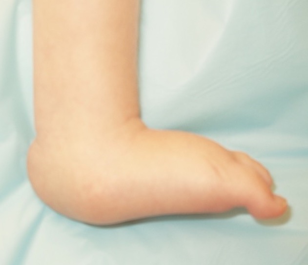
Clinical Features
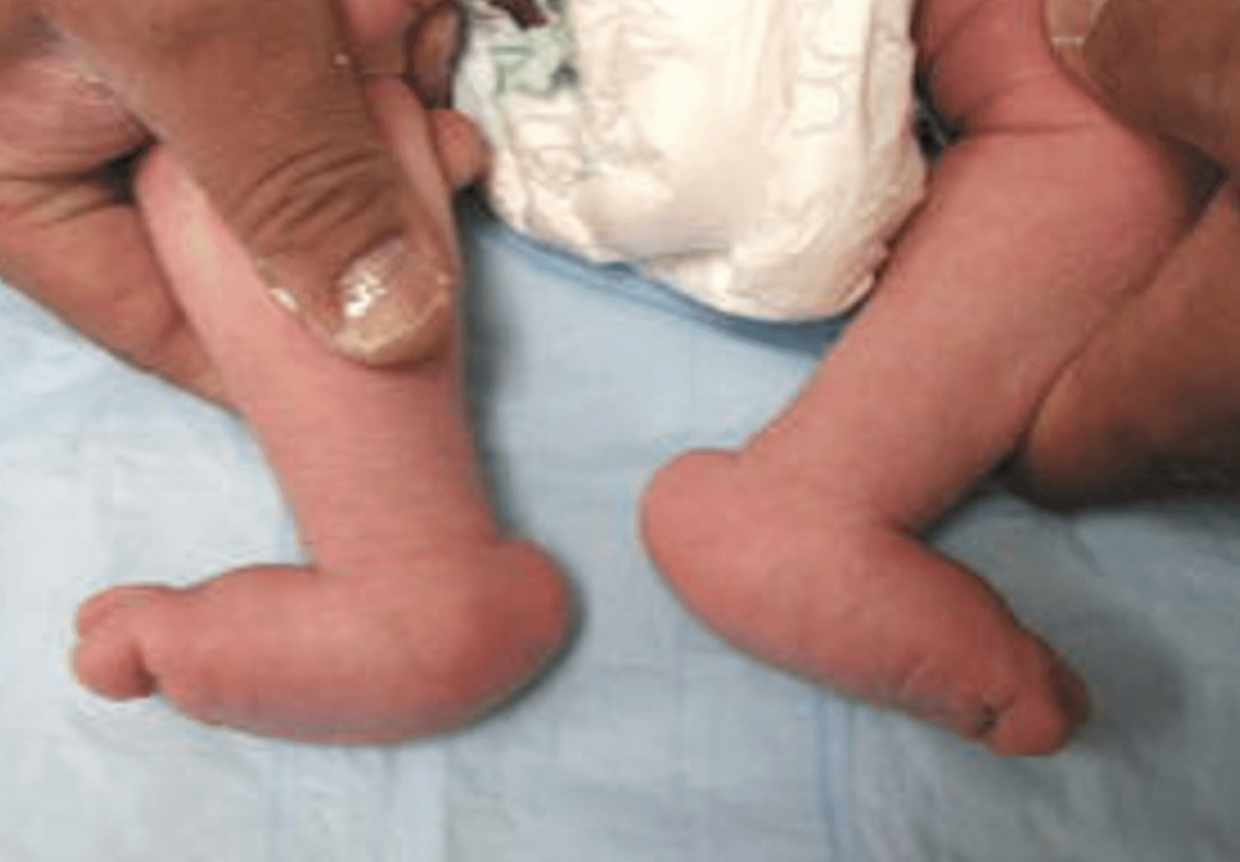
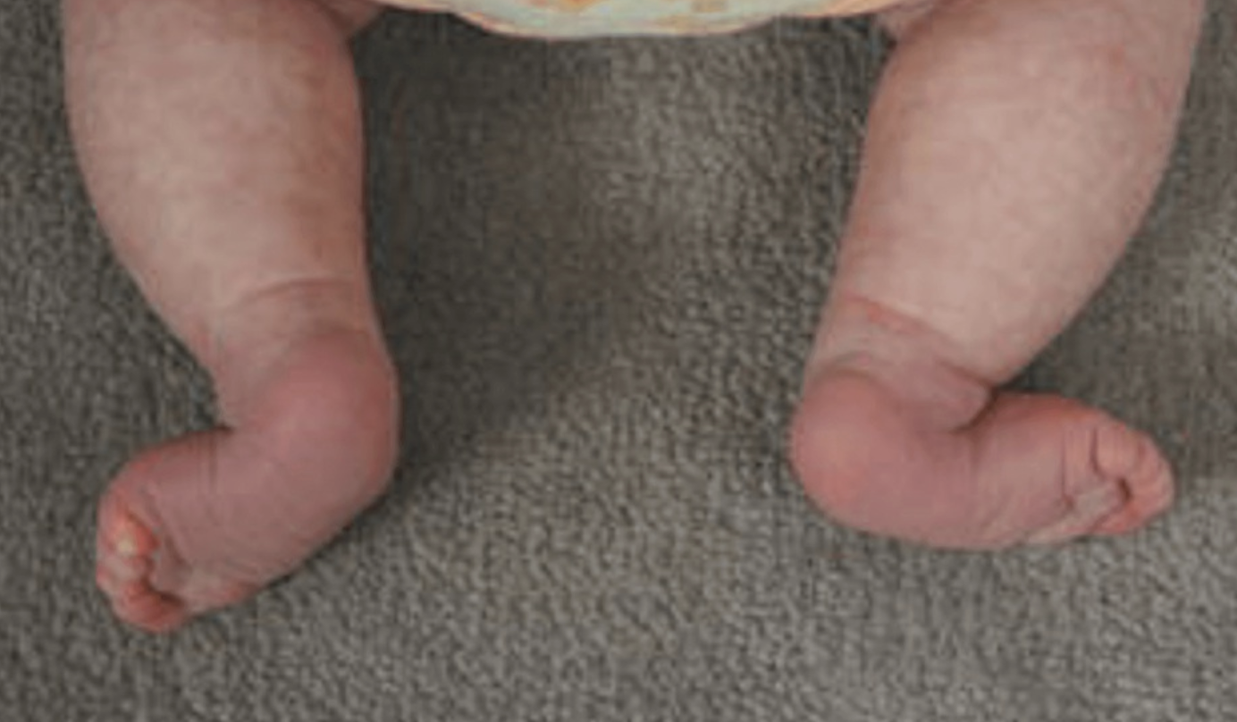
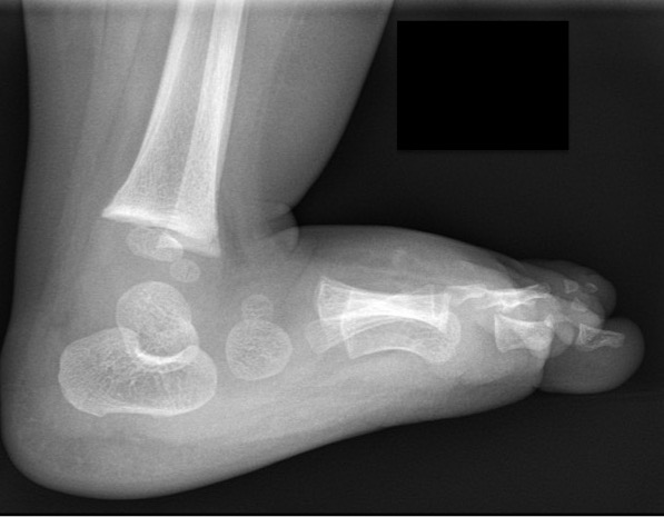
Images from: Alaee et al J Child Orthop 2007
Plantar aspect of foot convex / rocker-bottom appearance
Hindfoot - heel in fixed equinus
Forefoot - dorsiflexed and abducted
Differential diagnosis
Positional calcaneovalgus - flexible deformity with normal xrays
Posteromedial tibial bowing with calcaneovalgus foot
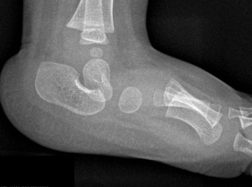
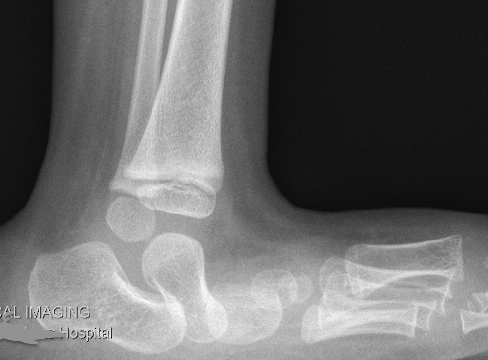
Xray
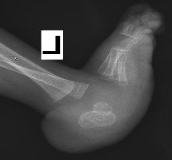
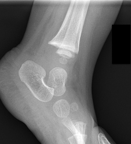
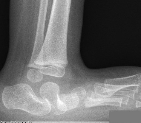
Lateral xray
- talus vertical
- calcaneum equinus
- increased talo-calcaneal angle
- talonavicular joint dislocated
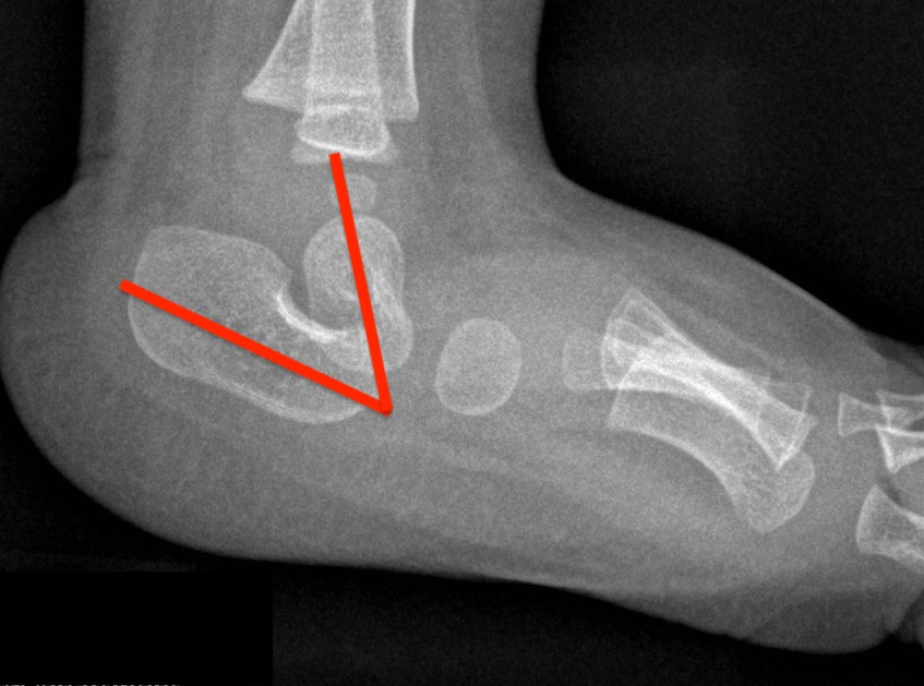
Increased talo-calcaneal angle in CVT
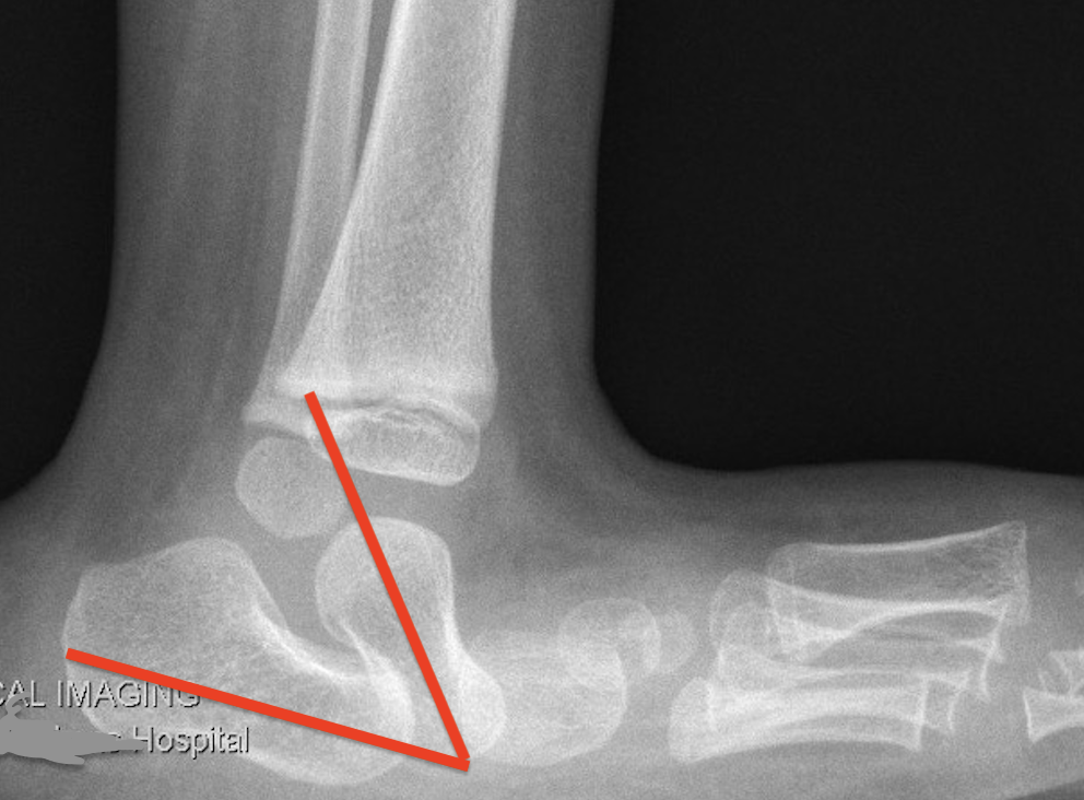
Increased talo-calcaneal angle with dislocated talonavicular joint
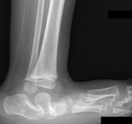
Dorsiflexion / plantarflexion views
- maximum dorsiflexion view - fixed equinus, talus still vertical
- maximum plantarflexion view - irreducibility of midfoot onto hindfoot
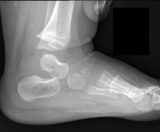
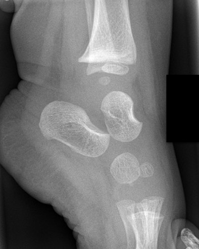
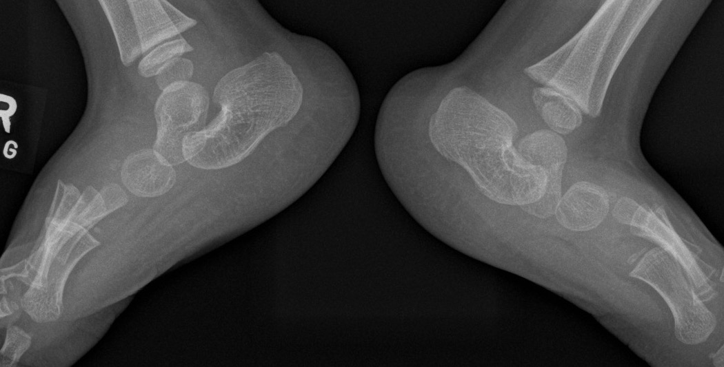
Normal foot in dorsiflexion / plantarflexion
Congenital vertical talus in dorsiflexion / plantarflexion
Management
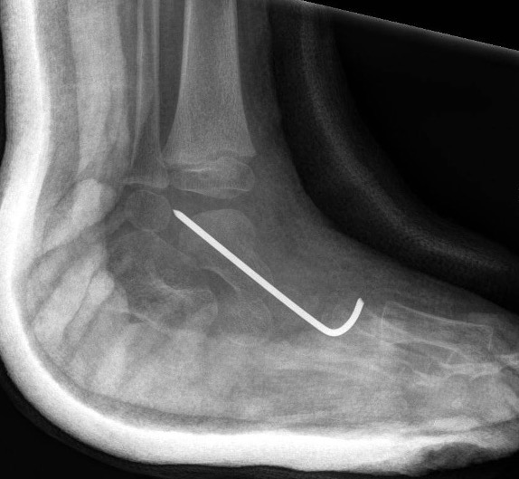
Dobbs Method - casting + minimally invasive surgery + bracing
Bouchard Journal of POSNA 2022 PDF
1. Reverse Ponseti technique
- weekly casting beginning week 1
2. Minimally invasive surgery
- typically 9 months of age due to risks of GA
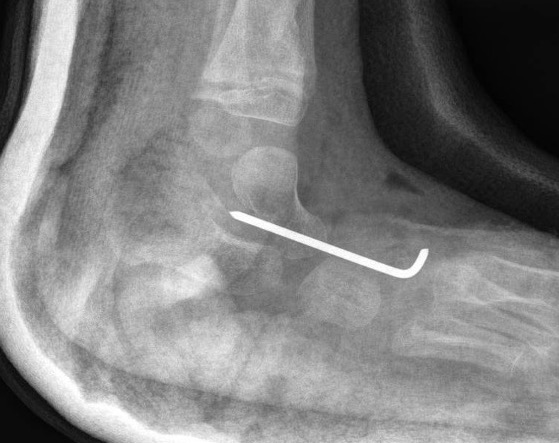
- reduce and K wire talonavicular joint +/- lengthen tibialis posterior
- percutaneous Achilles tenotomy
- +/- dorsal extensor tendon lengthening if needed
3. Brace / cast
- AFO full time for 3 months
- night bracing for 2 eyars
Open reduction
Indication
Severe, non responsive CVT
Technique
Approach dorsal / posterior
1. Reduction talonavicular joint
- release tibialis anterior and capsule
- reduce and K wire joint
2. Release forefoot dorsiflexion
- lengthen toe extensors and peroneals
3. Release hindfoot equinus
- T Achilles lengthening
- posterior capsulotomy ankle and subtalar joint
+/- naviculectomy
Results
Approach
Cummings et al J Pediatr Orthop 2023
- systematic review of surgery for CVT
- overall recurrence of talonavicular dislocation 19%
- recurrence direct medial approach: 29%
- recurrence dorsal approach: 11%
- best clinical score with Dobbs method
Minimally invasive
- 42 CVT feet
- extensive soft tissue release versus minimally invasive release
- better pain scores and ROM with minimally invasive Dobbs method
Late presenting
Triple arthrodesis