Definition
Degenerative arthritis at trapeziometacarpal joint (CMC)
- trapezoid - metacarpal
Epidemiology
Commonest hand joint involved in OA
Most common in older women
- 90% are females > 50 years
- asymptomatic degenerative changes common
Associated with arthritis in scapho-trapezial joint in 50%
Anatomy
1. Trapeziometacarpal Joint (TMJ)
2. Scaphotrapezial Joint (STJ)
3. Trapeziotrapezoidal Joint
4. Trapezium - Index Metacarpal Joint
The last two joints are rarely involved with OA
Saddle shaped
- allows movement in 3 planes
- flexion / extension
- adduction / abduction
- opposition
Volar, palmar oblique "beak" ligament
- provides stability of TMT
- origin is volar tubercle trapezium
- insertion ulna base of MC
- resists dorsal subluxation
Palmar 1/2 loaded > dorsal
- 13 x pressure with pinch
Aetiology
Primary
Combination of
- high compressive loads
- relatively unstable joint
- complex range of movement
May be related to ligamentous laxity
Secondary
Gout
Rheumatoid arthritis
Infection
Trauma
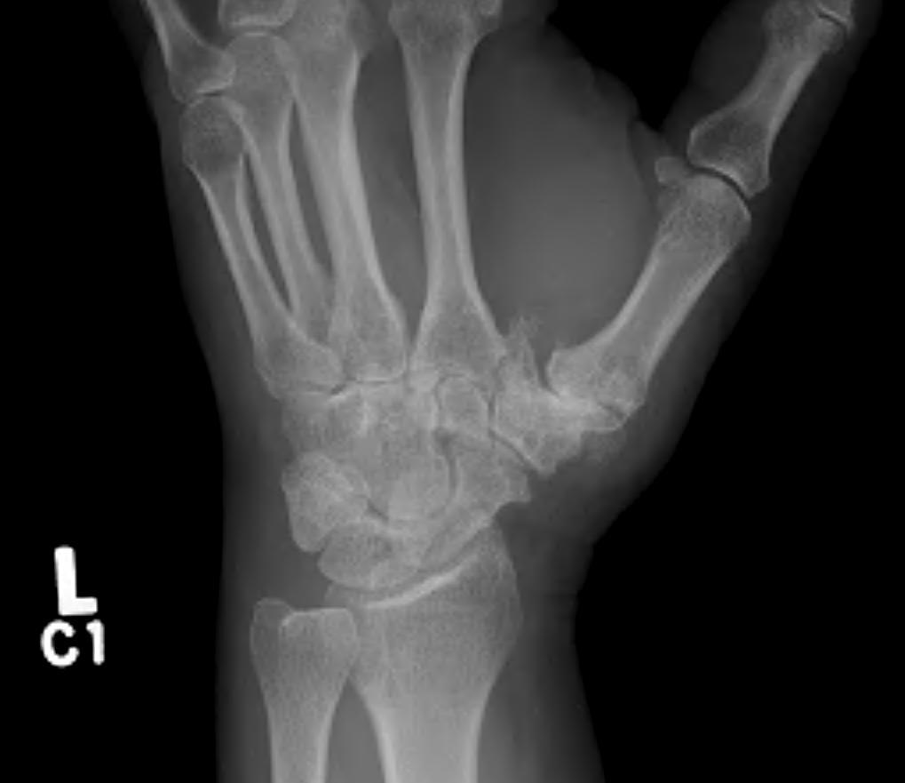
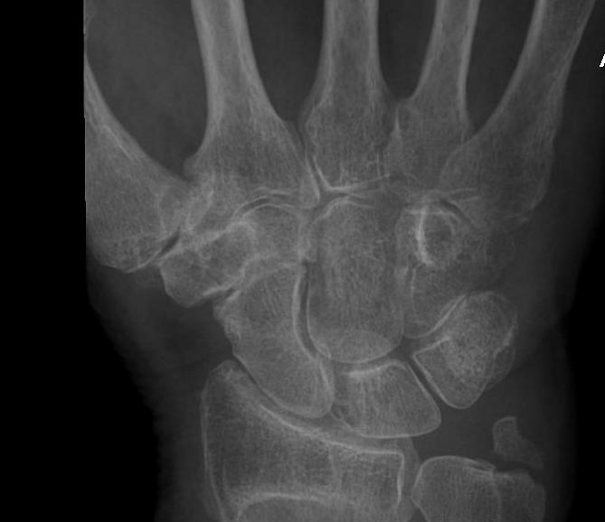
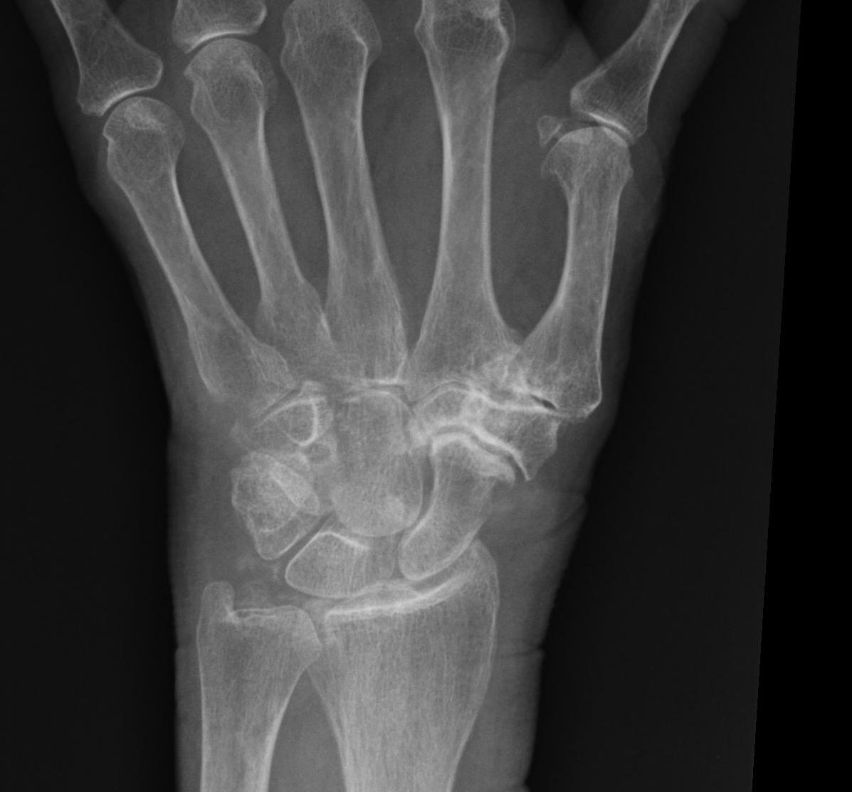
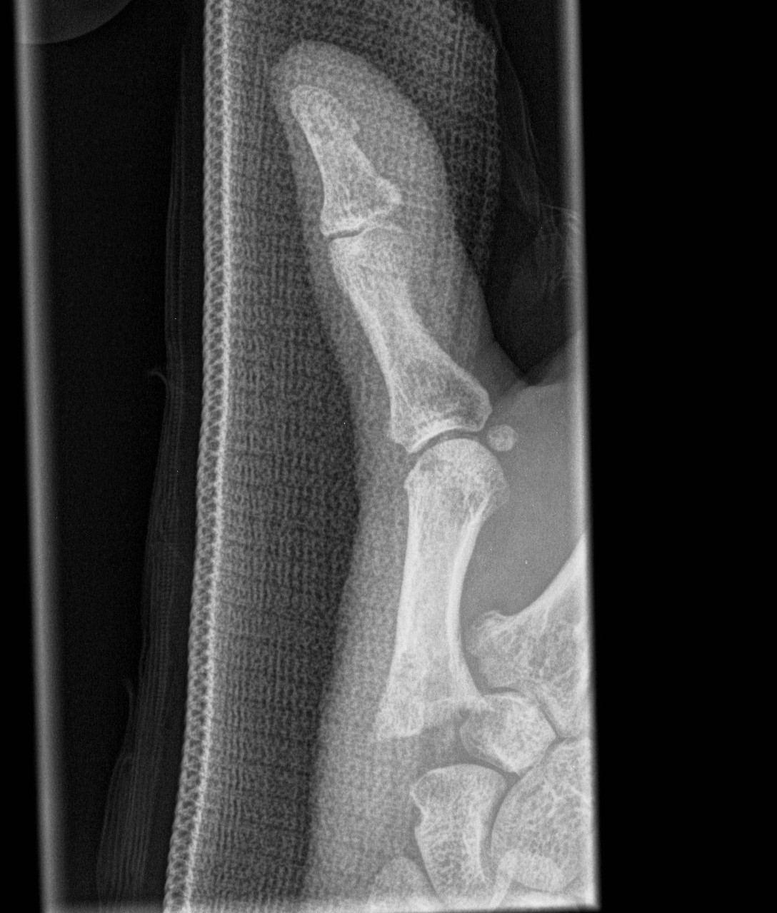
Eaton Classification
Stage 1
Joint normal with synovitis
Stage 2
Joint space narrowed
- may be mild subluxation (< 1/3)
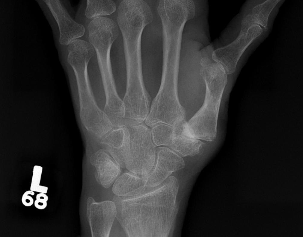
Stage 3
Joint space obliterated
Subluxation base of thumb
- adducted position
- proximally is anchored by adductor pollicis
- base subluxes radially / beak ligament ruptured
Stage 4
Involvement of multiple joint surfaces especially STT joint
Symptoms
Pain at base of thumb especially with pinch grip
Becomes constant / difficulties with ADL
Stiff thumb
Weak pinch grip
Examination
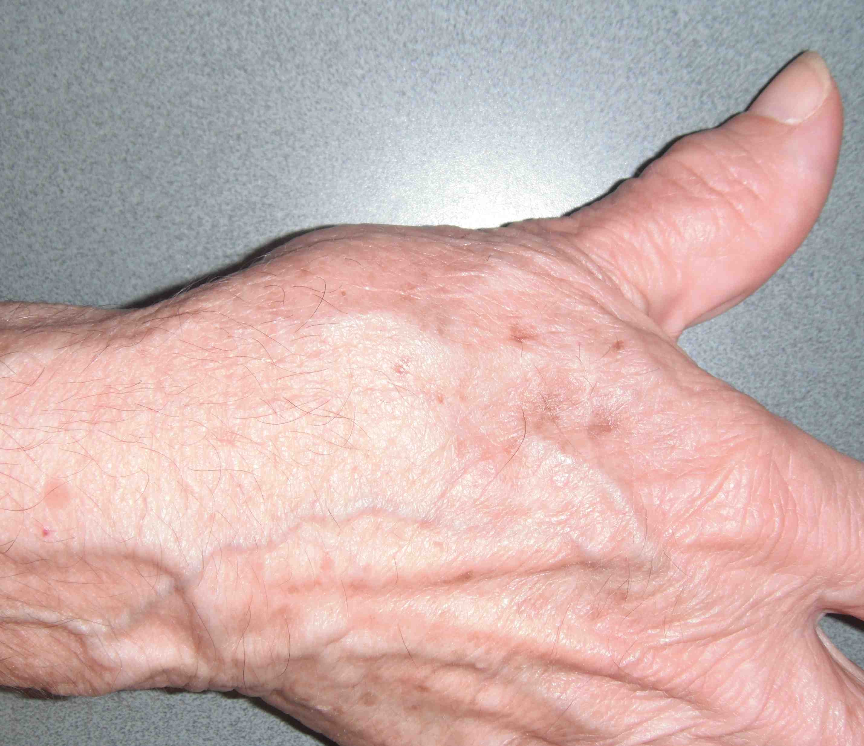
Tenderness around CMC joint
Swelling from
- synovitis
- osteophytes
Positive grind test
- passive thumb circumduction and axial loading
- causes pain
Web space contracture
- fixed flexion-adduction contracture of 1st MC
- compensatory MCPJ extension
DDx
De Quervain's
Radiocarpal OA
SNAC / SLAC
Scaphoid nonunion
Carpal tunnel syndrome
FCR synovitis
Volar ganglion
SRN neuroma
Management
Nonoperative Management
Majority of patients do not require surgery
Options
Rest / static splinting / thumb spica
Oral analgesics and NSAIDS
Intra-articular steroids / US guided
Operative Management
1. Reconstruction of the volar ligament
Indication
- stage 1 disease
- non responsive to non operative management
Advantages
- minimises progression of degenerative changes
Technique
- reconstruction of the volar ligament with slip FCR
- tendon passed through MC base and trapezium
- create stabilising ligament (tenodesis)
2. CMC Arthrodesis
Indication
- stage II and III disease
- young manual workers
- ligamentous laxity and neurological conditions
Contraindications
- pantrapezial OA
- i.e. involvement of STJ
Advantages
- pain-free
- strong pinch
- allows heavy use
Disadvantages
1. Limits mobility of thumb MC
- loss of abduction / adduction
- unable to put palm flat on table
2. Increases stress on adjacent joints
Position
- thumb position when fist made
- 30-40o palmar abduction
- 10-15o radial abduction
Technique
- dorsal incision at base of thumb over CMCJ
- dorsal to APL, between EPL and EPB
- protect SRN
- protect radial artery as it passes dorsally over STJ
- transverse incision capsule
- cut articular surfaces with saw
- ensure can pinch grip with IF / MF
- ensure can place across palm
- headless compression screws / plate
- POP for 6 weeks
3. Hemitrapeziectomy
Removal of distal half of trapezium only
4. Excisional arthroplasty / trapeziectomy
Indications
- stage II & III disease
- no significant MC subluxation
Technique
- simple excision of trapezium
Advantages
- simple procedure
- minimal immobilisation
Disadvantages
- shortening of thumb ray
- weakness of pinch
- thumb adduction
Results
- trapeziectomy without interposition / ligament reconstruction
- no evidence has worse results than any other more complicated procedure
5. Trapeziectomy and LRTI
Indications
- stage III and IV disease
Concept
- trapeziectomy +
- ligament reconstruction of beak ligament with FCR / PL
- tendon interposition (FCR / PL / Capsule)
Supposed Advantages
- maintains strength / pinch grip
- prevents shortening
Disadvantages
- tendon harvest
- longer / more involved procedure
- no evidence of improvement of pinch grip / prevention of shortening
Approach
Incision
- dorsoradial
- junction of volar and dorsal skin
Dissection
- protect SRN
- between APL and opponens
- radial artery over ST Joint
- open capsule over trapezium
- elevate thenar muscles from trapezium and 1st MC
Excise trapezium
- remove bone piecemeal / or in one piece
- take care not to damage underlying FCR
LRTI Technique 1
Make hole in base of MC
- perpendicular to plane of thumbnail
- from radial cortex to base
Harvest lateral half FCR
- 10 - 12 cm strip
- 2 - 3 transverse incisions in forearm over FRC
- split all the way to base of second MT
- pass through base second MT then radial cortex
- pass around base to resurface
- suture to itself whilst pushing MC base medially
Make spacer
- anchovy tendon on itself
- insert it into trapezium fossa
Stabilise with K wire
- MC reduced and out to length
Close wound & apply thumb spica
Postoperative
- ROS and K wire at 10 days
- splint for another 3 weeks
- progressive exercises
LRTI Technique 2
Harvest PL
- leave attached distally
- pass into base of thumb under FCR to where trapezium used to be
- ligament suspension by passing through radial capsule and FCR multiple times
- tightens the capsule and FCR into the gap
Capsular interposition technique
Open capsule as a distally base flap
- after trapeziectomy suture into base of wound as interposition
Results
- > 90% satisfactory results long-term
- > 95% pain relief
- > 90% increased grip strength
- average loss of height is 13% at 9 years
6. Silicone replacement arthroplasty
Indications
- stage III and IV disease
- low-demand patient
- rheumatoid
Concept
- trapeziectomy
- insert silicone trapezium
Advantages
- retains movement at CMC joint
Disadvantages
- subluxation or dislocation
- prosthesis breakage (50% at 4 years)
- silicone synovitis
Issue
- address subluxation by soft tissue reconstruction
- strip of APL can be passed through hole in prosthesis
7. Joint replacement
High revision rate
- pain
- lysis
- loosening