Hoffa fracture
Definition
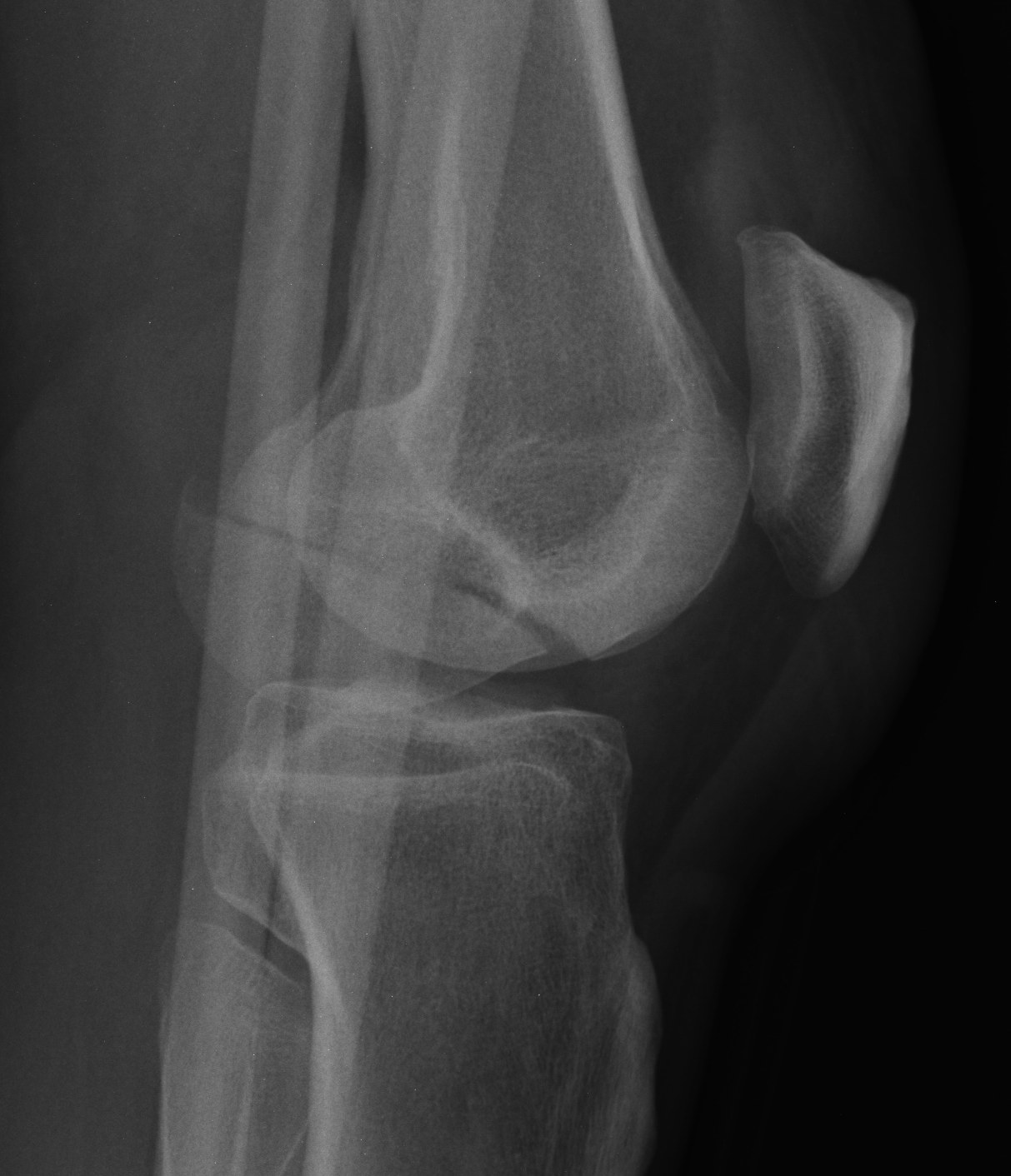
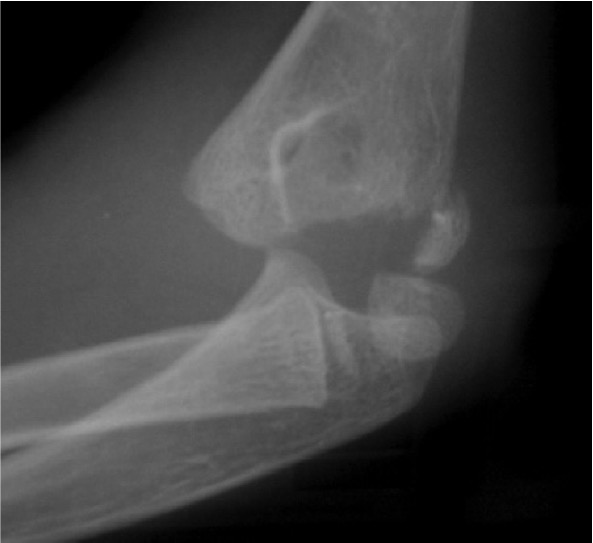
Coronal plane fracture of distal femoral condyle
- intra-articular
- often only attachment is posterior capsule
Epidemiology
Rare
Mechanism
Usually a severe valgus trauma
Xray
Coronal plane fracture of distal femoral condyle
- intra-articular
- often only attachment is posterior capsule
Rare
Usually a severe valgus trauma
Insufficiency fracture
- secondary to exceeding fatigue threshold
- usually of second or third MT shaft
Onset of new and very intense / strenuous physical activity
- i.e. new army recruits / dancers
Women with postmenopausal osteoporosis
Cavus feet
Children < 6
- entire distal humerus physis is displaced
Distal physis not ossified < 1 year
- may be a difficult diagnosis
Uncommon
< 1% Primary bone tumour
Young boys
- second decade
Similar to OO
Spine 30%
- especially posterior elements
Long bones 35%
Back or limb pain
- pain less severe than OO
Adult form
- 45 year old females
- more severe than Kohler's
Intense pain +/- oedema & inflammation
- often pronounced limp
- marked flat foot with prominant navicular
Navicular narrowed
- lateral part dense, sclerotic & thin
- occasional fracture line
Congenital Talipes Equinovarus
Congenital abnormality of the foot characterised by
- hindfoot equinus & varus
- forefoot Adduction
- midfoot Supination
Latin: talus - ankle / pes - foot / equinus - horse like
Foot
- pipe stem calf
- short wide foot
- small heel
- curved lateral border
Fracture distal to articular surface & proximal to intertrochanteric region
On average 4 years younger than intertrochanteric fracture
One year mortality as high as 36%
Only 1/3 will return to pre-fracture living environment
Bilateral Pars Fracture C2
- traumatic axis spondylolisthesis
Neurological injury uncommon
- fragments separate and decompress
Different to judicial hanging where spinal cord is severed
Rare
- unilateral
- bilateral
Compression
Lateral Compression
Rotation
Skull base pain
Cock Robin
Cranial nerve injury
Type I
Impaction of a condyle
Facet joint dislocations secondary flexion distraction injury
10%
1. Unifacet subluxation - interspinous process widening
2. Unifacet dislocation - 25% anterolisthesis
3. Bifacet dislocation - 50% anterolisthesis
4. Complete vertebral translation - 100% anterolisthesis