Cord injury patterns
Anatomy
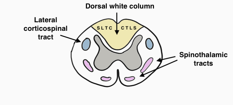
Dorsal Columns
- light touch, vibration & proprioception
- CTLS (cervical fibres central, sacral fibres lateral)
Lateral Corticospinal Tract
- motor tract
Dorsal Columns
- light touch, vibration & proprioception
- CTLS (cervical fibres central, sacral fibres lateral)
Lateral Corticospinal Tract
- motor tract
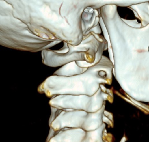
Acute dislocation of the atlanto-axial facet joint
Present with acute torticollis
Children
Injury to ulnar collateral ligament of thumb MCPJ
Interferes with pinch grip and grasp and thumb is ineffective as a post

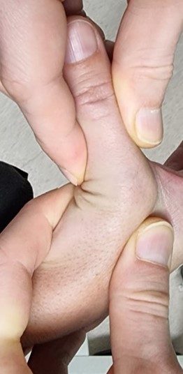
Middle age men
Steroids / Growth Hormone
Usually occurs in gym
Bench Press
Significant bruising in the acute phase
In chronic setting, ask patient to adduct against hip / resistance
Origin
- on right arises from brachiocephalic trunk behind right sternoclavicular joint
- on left arises from the arch of the aorta
Ends
- outer border first rib as axillary artery
First part
- origin of artery to medial aspect scalenus anterior
- on right arches above the clavicle / lies on the pleura / RLN wraps about it
Enters thigh
- midway between ASIS and pubic symphysis
- in femoral triangle
- NAVY (nerve / artery / vein / Y fronts)
- in femoral sheath with femoral vein (transversalis fascia and psoas fascia)
- femoral nerve outside sheath / under the iliac fascia / lateral
Femoral triangle
Anatomy
- inguinal ligament superiorly
- medial border sartorius laterally
Benign enlargement of the common digital branch
- usually 3rd webspace
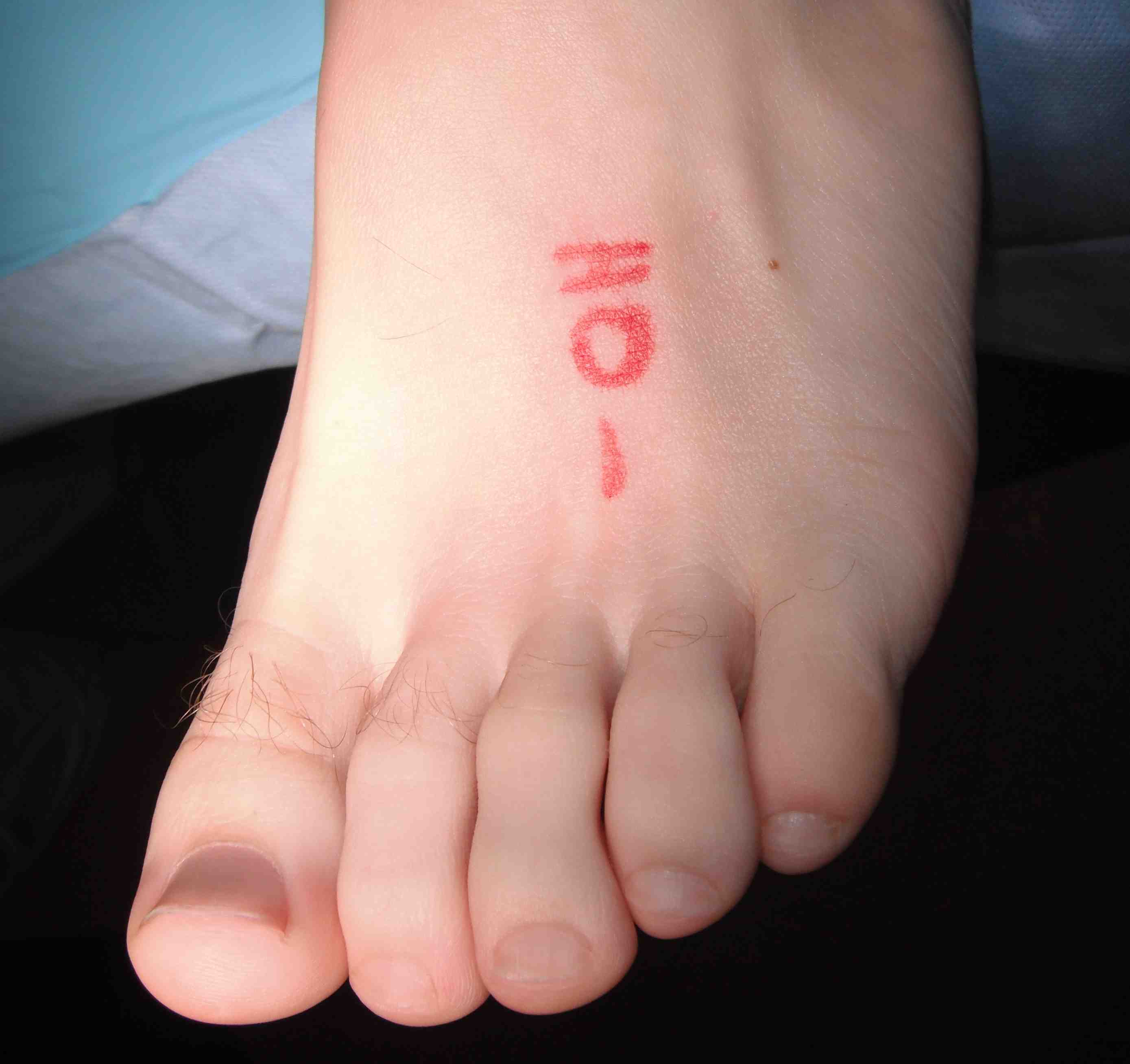
Found at level of or just distal to MT heads
- deep to the deep transverse MT ligament
Classically women between 40 and 60
Fascicles of long, spiraling bundles
- tenocytes & Type I collagen
- synovial cells & fibroblasts present
Endotenon
- surrounds the individual collagen bundles
Epitenon
- fine fibrous outer layer, highly cellular, continuous with endotenon
- contains most of the blood vessels & capillaries
5 Annular pulleys
3 Cruciate pulleys
A1 and A5 expendable
Loss of other annular pulleys can lead to bowstringing
- A2 & A4 +/- A3
Rock climbers
- usually when slipping
May hear or feel a pop
Develop swelling / tenderness / pain
Bowstringing
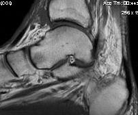
Osseous canal between talus and calcaneum
- interosseous talo-calcaneal ligament
- cervical ligament
- joint capsule
- nerve endings / arterial anastomoses
Flat foot / overpronation
Inversion / sprain