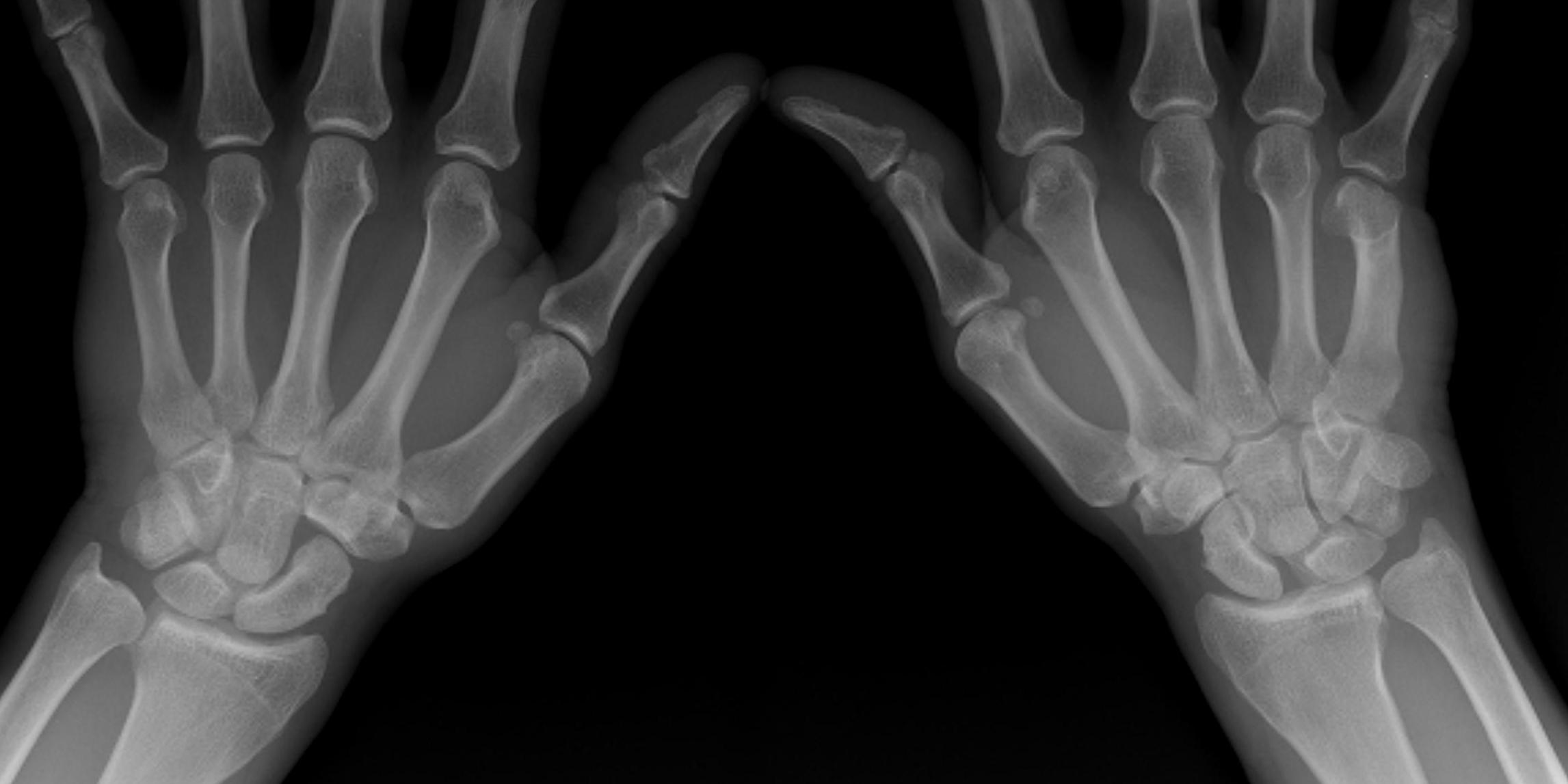
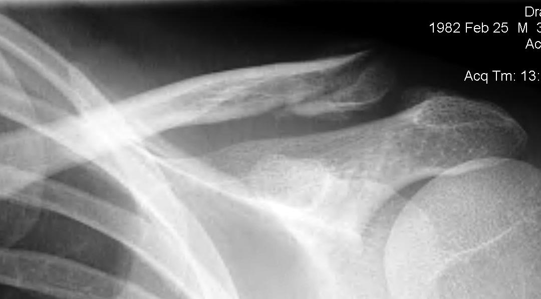
surgery

Facet Joint Dislocation

Definition


Facet joint dislocations secondary flexion distraction injury


Epidemiology


10%


Stages


1. Unifacet subluxation - interspinous process widening

2. Unifacet dislocation - 25% anterolisthesis

3. Bifacet dislocation - 50% anterolisthesis

4. Complete vertebral translation - 100% anterolisthesis


Unilateral Facet Joint Dislocation


Morton's Neuroma

Definition


Benign enlargement of the common digital branch

- usually 3rd webspace


Mortons Neuroma Common Site


Anatomy


Found at level of or just distal to MT heads

- deep to the deep transverse MT ligament


Epidemiology


Classically women between 40 and 60

Great toe dislocation

Epidemiology


Uncommon

- dancers

- athletes


Aetiology


Hyperdorsiflexion of the MTPJ


Pathology


MT head dislocates plantar

- may buttonhole through capsule

- can prevent closed reduction


Blocks to Reduction


1.  Sesamoids

2.  Conjoint tendon

3.  Intersesamoid Ligament


TSA Technique

TSR APTSR Lateral


Goal

- head that translates 50% in all directions

- allows 30o ER with arm at side

- stable posteriorly


Pre-Op 

- antibiotics

- consider specific Propionobacterium cover

Surgery

Indications 


1. Significant functional impairment


2. PIPJ contracture

- originally thought to intervene early

- Macfarlane showed residual FFD always about 30o

- may need to release  check rein ligaments / accessory collateral ligaments


3. MCPJ contracture >30o


4. Trigger fingers

- must do limited fasciectomy 

Management

Operative v Nonoperative Literature


1.  Buckley etal JBJS Am 2002


Prospective multi-centred RCT

- 309 displaced intra-articular fractures

- operative v non operative management

- 2 year follow up


Findings

- used patient orientated functional outcomes

- overall VAS and SF36 not significantly different between 2 groups


Improved Operative Outcome if

- not workers compensation

- women

- < 29

Heterotopic ossification

Definition


Pathological bone formation in soft tissues


Epidemiology


In elbow

- 3% of trauma

- 89% if head injury + trauma


Types


Completely different


1.  Myosisitis Ossificans Circumscripta

- post traumatic

- more common

- recognised as a consequence of neurological injury


Synovial Chondromatosis

DefinitionSynovial Chondromatosis Knee MRI


Chondroid Metaplasia of synovium affecting large joints


Nodules of hyaline cartilage

- formed in the subsynovial layer of joint capsules


Epidemiology


Rare lesion

Most common in 20's and 30's

Sex: M > F (2:1)

Monoarticular


Site


Bipartite Patella

Ossification


Patella may develop from one or multiple ossification centres at 3 years


Failure of centres to fuse may produce bipartite or tripartite patella

- usually bilateral and painless


Classically superolateral


Classification Saupe


I   Inferior Pole 5%

II  Lateral 20%