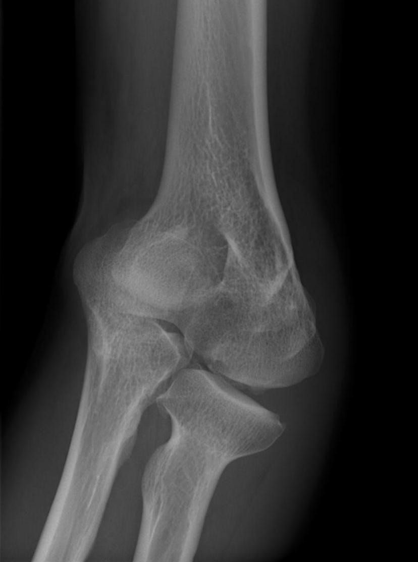
operative management

Lateral Epicondylitis / Tennis Elbow

Incidence


Lateral : Medial 9:1


Epidemiology


4th & 5th decades

- M = F

- 75% dominant arm 


50% of regular tennis players

- especially > 2 hrs / week


Aetiology


Insertion pathology / Enthesopathy


Over-extension of the elbow with supination / pronation


Anatomy


Lateral epicondyle

- anconeus from posterior face

- ECRB and EDC from anterior face (CEO)

Cubitus Varus

Incidence


Occurs 10% of supracondylar humeral fractures

- varus malunion


Lateral condyle fracture

- AVN trochlea


Elbow Cubitus Varus


Growth arrest of medial aspect physis

- rare

- post traumatic


Effects


Achilles Tendonopathy

Definition


Inflammation of achilles tendon; insertional or noninsertional


Spectrum


Tendonitis / Tendonosis / Rupture


Anatomy


Triceps surae

- medial and lateral gastrocnemius

- soleus

- surrounded by paratenon which allows gliding and supplies nutrition


Inserts middle 1/3 calcaneal tuberosity

- 2 x 2 cm area

- 90o rotation distally


Retrocalcaneal bursa (x2)

Achilles Tendon Rupture

AnatomyAchilles tendon anatomy


Gastrocnemius tendon 10-25 cm long

- soleus 3-10 cm

- inserts superior calcaneal tuberosity

- fibres spiral 90°

- fibres that lie medially in proximal portion become posterior distally

- allows elastic recoil & energy storage


Plantaris present in 90% population

- medial to T Achilles


Poor blood supply midportion

- mesotenal vessels

Tendonitis / Tendonopathy

Anatomy


Lateral compartment of leg

- run through retromalleolar groove

- pass superior and inferior to peroneal tubercle

- covered by inferior peroneal retinaculum


Peroneus longus

- origin lateral condyle of tibia and head fibula

- tendon PL superficial and inferior to brevis in retromalleolar groove

- runs in cuboid groove 

- insert plantar surface base of 1st MT and lateral aspect medial cuneiform

Spinal Metastasis

EpidemiologyThoracic Metastasis


80% cancer patients have spinal metastasis at autopsy 


Spine is number one site for bony metastasis (50%)


Cause


Hexagon: PBBLTK


                 Prostate    Breast


Scheuermann's kyphosis

Definition 


Structural kyphosis of thoracic or thoracolumbar spine

- characterised by vertebral wedging & subsequent growth disturbance of vertebral end plate


X-ray Diagnostic Criteria Sorensen 1964


1.  Kyphosis > 45°


2. > 5° wedging 3 or more adjacent apical vertebrae


Other features

- Schmorl nodes

Vertebral Osteomyelitis

Epidemiology


M:F =2:1


30-40 years


20% diabetic


50-80% identifiable source


Site


Lumbar (50%) > Thoracic > Cervical (<10%)


Pathogenesis


1.  Haematogenous

- arterial rather than venous


Risk factors

- UTI (40% of all cases)

- IVDU

- elderly