non operative management

Plantar Fasciitis

Definition


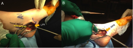
Pain at attachment of thickened central part of plantar aponeurosis to Medial Calcaneal Tuberosity


Anatomy Plantar Fascia


Origin 

- medial calcaneal tuberosity


Inserts 

- 5 bands superfical & deep layers


Superficial

- insert transverse MT ligament & skin


Deep 

- flexor sheath, volar plate & periosteum of P1


Tendonitis / Tendonopathy

Anatomy


Lateral compartment of leg

- run through retromalleolar groove

- pass superior and inferior to peroneal tubercle

- covered by inferior peroneal retinaculum


Peroneus longus

- origin lateral condyle of tibia and head fibula

- tendon PL superficial and inferior to brevis in retromalleolar groove

- runs in cuboid groove 

- insert plantar surface base of 1st MT and lateral aspect medial cuneiform

Dislocation

Definition


Anterior displacement of peroneal tendons out of peroneal groove


Epidemiology


Most common in young adults


Acute injury often missed 


Aetiology


Congenital


3 % neonates

- resolves spontaneously


Traumatic  


Occurs following sporting activities

Freiberg's

Definition


Crushing osteochondritis of metatarsal head


Frieberg's


Epidemiology


Usually 2nd metatarsal (80%)

- occasionally third

- can occur in any


Age 10-15 years

- peak 15 year old girls

- F:M = 3:1

- occurs during the growth spurt at puberty


Bilateral in 6%


Aetiology


Lumbar Herniated Discs

Epidemiology


Sciatica > 2/52 1.6%


M:F = 1:1


Most common L4/5 

L5/S1 inherently stable 


Risk factors


Sedentary lifestyle

Smokers

Frequent driving

Heavy lifting 


Anatomy


Annulus Fibrosis

- circumferential, multilayered rim