Lateral Clavicle Fracture
Epidemiology
Elderly population
Less common in younger population
Lateral 1/3 Neer Classification
Type I
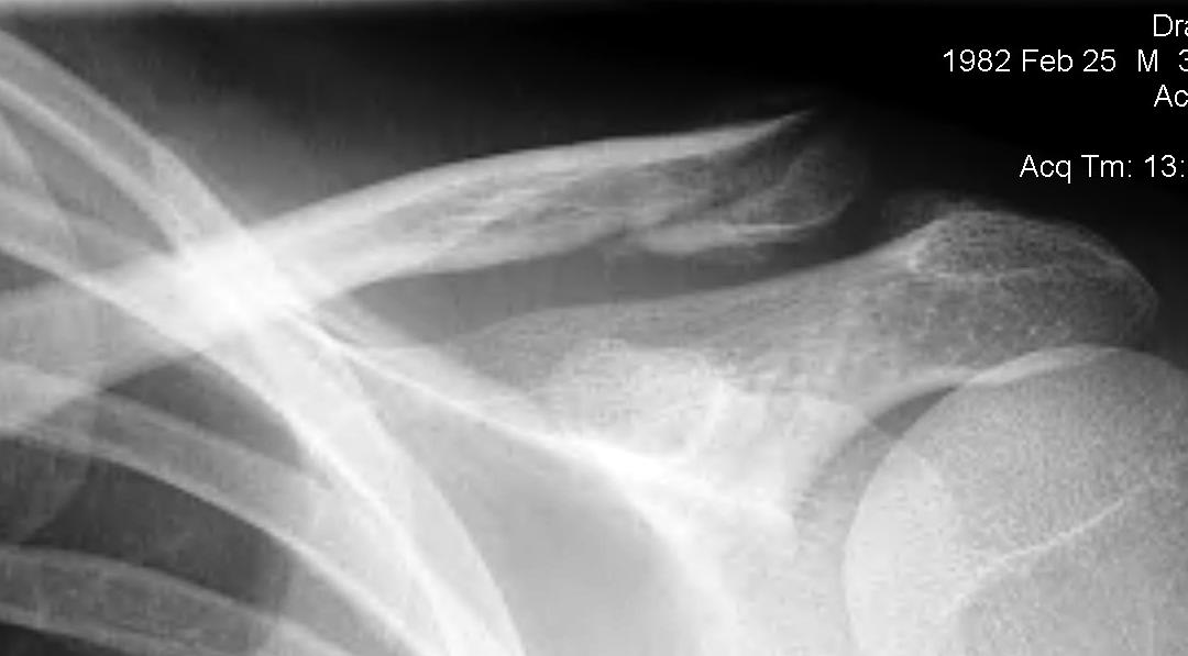
Fracture lateral to the CC ligaments
- does not extend into the ACJ
- non displaced
- most common 4:1
Elderly population
Less common in younger population
Type I
Fracture lateral to the CC ligaments
- does not extend into the ACJ
- non displaced
- most common 4:1
Chronic massive rotator cuff defect
- uncovered humeral articular cartilage
- high riding humeral head
- abrasion by undersurface of coracoacromial arch
Neer
- introduced term "cuff tear arthropathy"
- included significant rotator cuff diagnosis & arthritis in older patients
- especially women
- synovial fluid contained calcium phosphate crystals + proteases

Massive tear
1. > 5cm
- retracted to humerus / glenoid margin
2. At least 2 complete tendons
- lose SS / IS or SS / SC
Instability in at least 2 planes
- postero-inferior
- antero-inferior
- antero-postero-inferior
Recognised as a common problem
- often misdiagnosed
Most patients athletic
A GH dislocation which has been missed for a significant period of time
- time period is arbitary
- > 3-6 weeks
Humerus soft and osteoporotic
Significant soft tissue contractures
1. Anterior / subcoracoid dislocation
Beware
- scarring to NV structures
- RC tears including SSC, especially > 40
Most common form of shoulder instability
- young males
- M:F = 2:1
Indirect ER and abduction moment on arm
- disruption of anterior stabilisers
Initial injury
- severe pain in shoulder
Synovial joint with hyaline cartilage
Has fibrocartilage intra-articular disc
- complete or incomplete
- usually degeneration by 4th decade
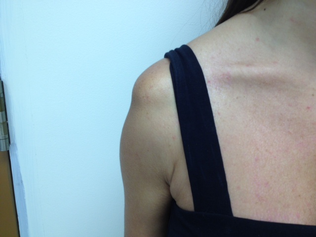
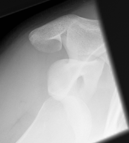
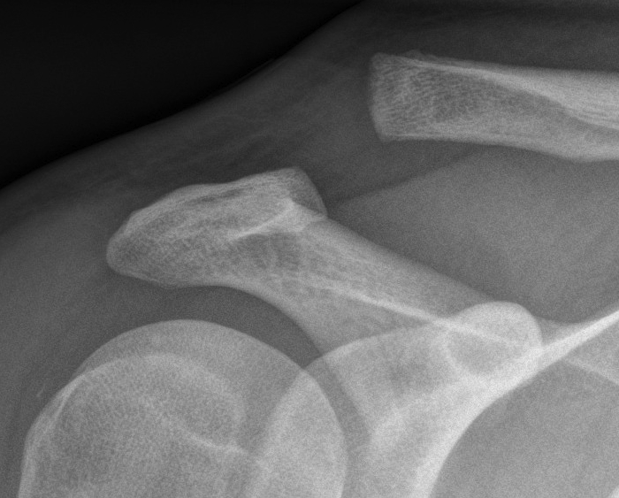
Clavicle may lie superior to acromion in normal population
Acromioclavicular Ligaments
Injury to ulnar collateral ligament of thumb MCPJ
Initial description
- chronic laxity of British gamekeeper's thumb's
- no specific trauma
- secondary to breaking pheasant's neck
Acute trauma
- snow ski
- ball games
Valgus / forced abduction
UCL
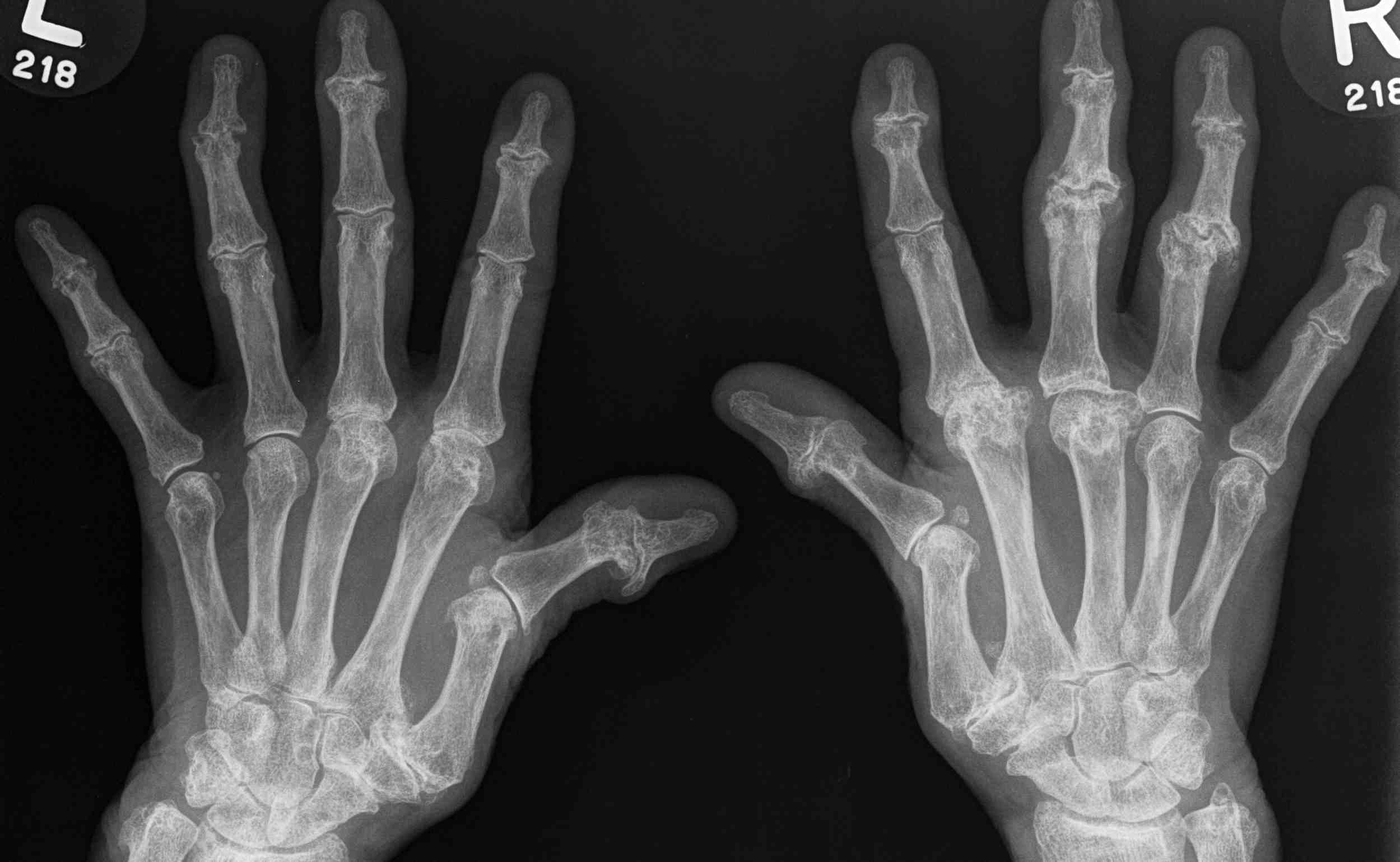
1987 American College of Rheumatology
Need 4/7 (MAX RANS)
1. Morning Stiffness
2. Arthritis of 3 areas > 6/52
3. Xray changes
4. Rh factor
5. Arthritis of Hand > 6/52
6. Nodules
7. Symmetric Arthritis > 6/52
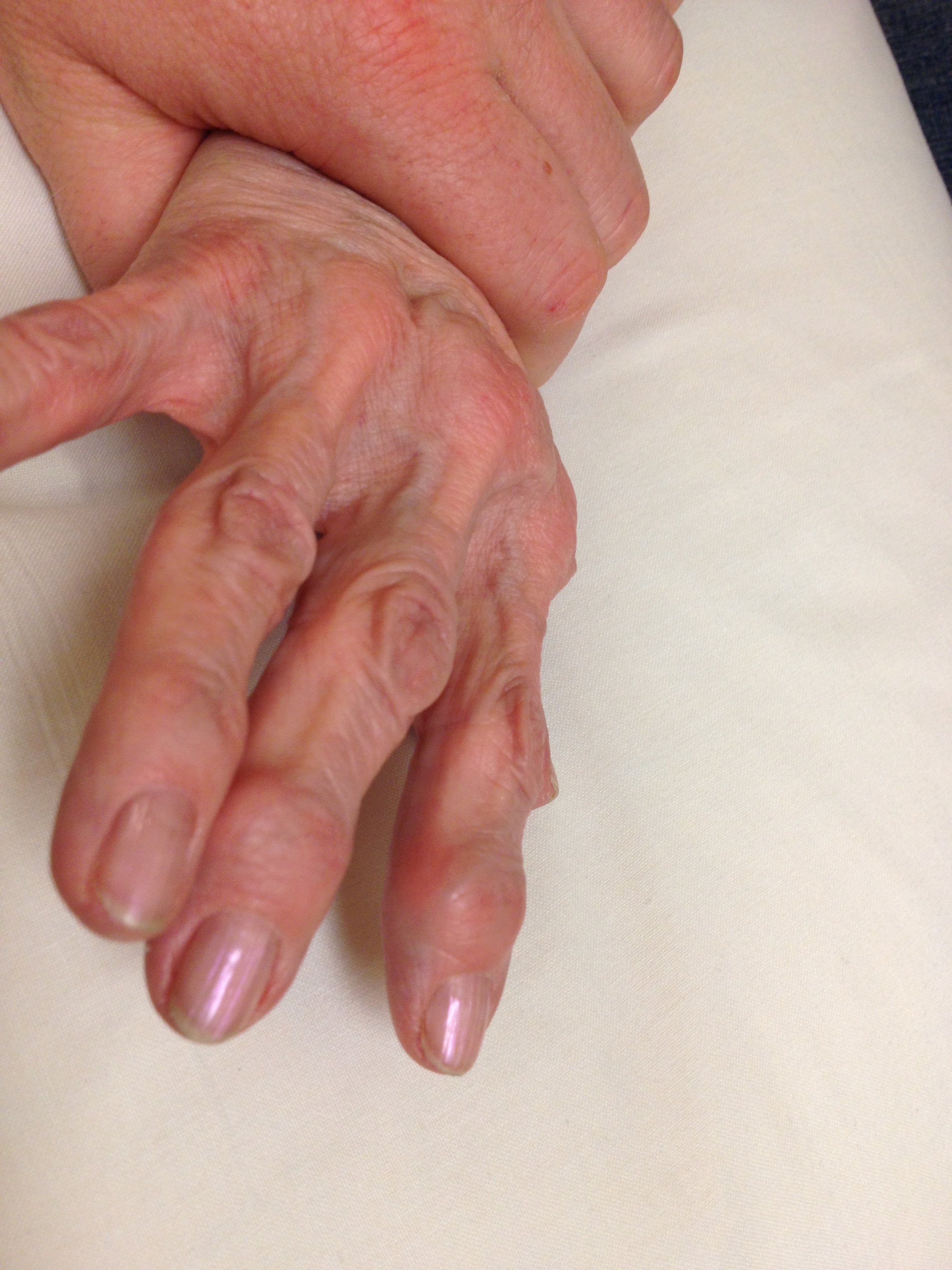
Male & Females > 60 years
- X-ray evidence of OA
Symptomatic
- 25% females
- 15% males
Base thumb
PIPJ / Bouchard's nodes
DIPJ / Heberden's nodes